
Eye Conditions treated in our Las Vegas Eye Practice
Refractive Errors
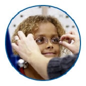
Refractive errors are the most frequent eye problems in the United States. Refractive errors include myopia (near-sightedness), hyperopia (farsightedness), astigmatism (distorted vision at all distances), and presbyopia that occurs between age 40–50 years (loss of the ability to focus up close, inability to read letters of the phone book, need to hold newspaper farther away to see clearly) can be corrected by eyeglasses, contact lenses, or in some cases surgery. The National Eye Institute states that proper refractive correction could improve vision among 150 million Americans.
Learn more about refractive errors
Age-Related Macular Degeneration

Macular degeneration, often called age-related macular degeneration (AMD), is an eye disorder associated with aging and results in damaging sharp and central vision. Central vision is needed for seeing objects clearly and for common daily tasks such as reading and driving. AMD affects the macula, the central part the retina that allows the eye to see fine details. There are two forms of AMD—wet and dry.
Wet AMD is when abnormal blood vessel behind the retina start to grow under the macula, ultimately leading to blood and fluid leakage. Bleeding, leaking, and scarring from these blood vessels cause damage and lead to rapid central vision loss. An early symptom of wet AMD is that straight lines appear wavy.
Dry AMD is when the macula thins overtime as part of aging process, gradually blurring central vision. The dry form is more common and accounts for 70–90% of cases of AMD and it progresses more slowly than the wet form. Over time, as less of the macula functions, central vision is gradually lost in the affected eye. Dry AMD generally affects both eyes. One of the most common early signs of dry AMD is drusen.
Drusen are tiny yellow or white deposits under the retina. They often are found in people aged 60 years and older. The presence of small drusen is normal and does not cause vision loss. However, the presence of large and more numerous drusen raises the risk of developing advanced dry AMD or wet AMD.
It is estimated that 1.8 million Americans aged 40 years and older are affected by AMD and an additional 7.3 million with large drusen are at substantial risk of developing AMD. The number of people with AMD is estimated to reach 2.95 million in 2020. AMD is the leading cause of permanent impairment of reading and fine or close-up vision among people aged 65 years and older.
Learn more about age-related macular degeneration
Cataract

Cataract is a clouding of the eye’s lens and is the leading cause of blindness worldwide, and the leading cause of vision loss in the United States. Cataracts can occur at any age because of a variety of causes, and can be present at birth. Although treatment for the removal of cataract is widely available, access barriers such as insurance coverage, treatment costs, patient choice, or lack of awareness prevent many people from receiving the proper treatment.
An estimated 20.5 million (17.2%) Americans aged 40 years and older have cataract in one or both eyes, and 6.1 million (5.1%) have had their lens removed operatively. The total number of people who have cataracts is estimated to increase to 30.1 million by 2020.
Diabetic Retinopathy

Diabetic retinopathy (DR) is a common complication of diabetes. It is the leading cause of blindness in American adults. It is characterized by progressive damage to the blood vessels of the retina, the light-sensitive tissue at the back of the eye that is necessary for good vision. DR progresses through four stages, mild nonproliferative retinopathy (microaneurysms), moderate nonproliferative retinopathy (blockage in some retinal vessels), severe nonproliferative retinopathy (more vessels are blocked leading to deprived retina from blood supply leading to growing new blood vessels), and proliferative retinopathy (most advanced stage). Diabetic retinopathy usually affects both eyes.
The risks of DR are reduced through disease management that includes good control of blood sugar, blood pressure, and lipid abnormalities. Early diagnosis of DR and timely treatment reduce the risk of vision loss; however, as many as 50% of patients are not getting their eyes examined or are diagnosed too late for treatment to be effective.
It is the leading cause of blindness among U.S. working-aged adults aged 20–74 years. An estimated 4.1 million and 899,000 Americans are affected by retinopathy and vision-threatening retinopathy, respectively.
Learn more about diabetic retinopathy
Glaucoma

Glaucoma is a group of diseases that can damage the eye’s optic nerve and result in vision loss and blindness. Glaucoma occurs when the normal fluid pressure inside the eyes slowly rises. However, recent findings now show that glaucoma can occur with normal eye pressure. With early treatment, you can often protect your eyes against serious vision loss.
There are two major categories “open angle” and “closed angle” glaucoma. Open angle, is a chronic condition that progress slowly over long period of time without the person noticing vision loss until the disease is very advanced, that is why it is called “sneak thief of sight.” Angle closure can appear suddenly and is painful. Visual loss can progress quickly; however, the pain and discomfort lead patients to seek medical attention before permanent damage occurs.
Amblyopia
Amblyopia, also referred to as “lazy eye,” is the most common cause of vision impairment in children. Amblyopia is the medical term used when the vision in one of the eyes is reduced because the eye and the brain are not working together properly. The eye itself looks normal, but it is not being used normally because the brain is favoring the other eye. Conditions leading to amblyopia include strabismus, an imbalance in the positioning of the two eyes; more nearsighted, farsighted, or astigmatic in one eye than the other eye, and rarely other eye conditions such as cataract.
Unless it is successfully treated in early childhood amblyopia usually persists into adulthood, and is the most common cause of permanent one-eye vision impairment among children and young and middle-aged adults. An estimated 2%–3% of the population suffer from amblyopia.
Strabismus
Strabismus involves an imbalance in the positioning of the two eyes. Strabismus can cause the eyes to cross in (esotropia) or turn out (exotropia). Strabismus is caused by a lack of coordination between the eyes. As a result, the eyes look in different directions and do not focus simultaneously on a single point. In most cases of strabismus in children, the cause is unknown. In more than half of these cases, the problem is present at or shortly after birth (congenital strabismus). When the two eyes fail to focus on the same image, there is reduced or absent depth perception and the brain may learn to ignore the input from one eye, causing permanent vision loss in that eye (one type of amblyopia).
Learn more about strabismus, just make an appointment
Other Eye and Eye Related Conditions
About Ophthalmology
Adie’s Pupil
Adie’s pupil, also known as tonic pupil, is a condition characterized by an abnormally large and sluggishly reacting pupil. It is caused by damage to the parasympathetic nerves that control the pupil’s constriction. This can result from an autoimmune disorder, viral infection, or trauma. Adie’s pupil typically affects one eye, causing it to appear larger than the other. The pupil may also be slow to react to light and have a characteristic “light-near dissociation,” where it constricts more with near vision than with bright light. While Adie’s pupil is usually benign, it can be a sign of an underlying neurological condition, so it’s important to have it evaluated by an eye care professional.
Adult Strabismus
Adult strabismus refers to the misalignment of the eyes in adults, which can cause double vision, eye strain, and other visual problems. It can develop due to a variety of factors, including eye muscle imbalances, nerve disorders, or previous eye surgery. Symptoms may include eye turning inward (esotropia), outward (exotropia), upward (hypertropia), or downward (hypotropia). Treatment options can include vision therapy, prism glasses, botox injections, or eye muscle surgery to realign the eyes. Proper diagnosis and management of adult strabismus is important to restore binocular vision and improve quality of life.
Age-Related Macular Degeneration
Age-related macular degeneration (AMD) is a leading cause of vision loss in older adults. It occurs when the macula, the central part of the retina responsible for sharp, straight-ahead vision, begins to deteriorate. There are two main types of AMD: dry AMD, which involves gradual vision loss, and wet AMD, which can cause rapid and severe vision loss due to abnormal blood vessel growth. Risk factors include age, smoking, family history, and certain genetic factors. While there is no cure, treatments like anti-VEGF injections and lifestyle changes can help slow the progression of wet AMD. Regular eye exams are crucial for early detection and management of this condition.
Here are the remaining paragraphs on the requested topics:
## Albinism
Albinism is a group of inherited genetic conditions that result in little or no production of the pigment melanin. This can affect eye, skin, and hair color. Ocular albinism primarily affects the eyes, while oculocutaneous albinism also impacts skin and hair. People with albinism often have very light-colored eyes, skin, and hair, and may be sensitive to bright light and glare. Vision problems are common, including reduced visual acuity, nystagmus (involuntary eye movements), strabismus, and photophobia. While there is no cure, low vision aids, tinted glasses, and sun protection can help manage symptoms. Genetic counseling is recommended for those with a family history of albinism.
## Alzheimer’s Disease, Dementia and the Eye
Alzheimer’s disease and other forms of dementia can impact various aspects of vision and eye health. Common visual changes include reduced contrast sensitivity, difficulty with depth perception, and problems with color vision. Patients may also experience difficulties with reading, recognizing faces, and navigating unfamiliar environments. Structural changes in the eye, such as cataracts and optic nerve degeneration, are more common in those with Alzheimer’s. Regular eye exams are important for early detection and management of vision problems associated with dementia. Caregivers should be aware of potential vision issues and make necessary accommodations to ensure the safety and quality of life for those affected.
## Amblyopia
Amblyopia, commonly known as “lazy eye,” is a condition in which one eye fails to develop normal visual acuity, even with corrective lenses. It is often caused by strabismus, refractive errors, or visual deprivation in early childhood. If left untreated, amblyopia can lead to permanent vision loss in the affected eye. Treatment typically involves correcting any underlying refractive errors, patching or blurring the stronger eye to force use of the weaker eye, and vision therapy exercises. Early detection and treatment, often before age 6, is crucial for restoring normal vision and preventing long-term visual impairment. Regular eye exams are recommended for children to identify and address amblyopia promptly.
## Anisocoria
Anisocoria is a condition characterized by unequal pupil size between the two eyes. It can be physiologic (normal variation) or pathologic (caused by an underlying condition). Pathologic anisocoria may be due to damage to the sympathetic or parasympathetic nerves that control pupil size, or it can be a sign of a serious neurological disorder, such as a brain aneurysm or tumor. Symptoms may include headache, eye pain, vision changes, or other neurological symptoms. Anisocoria should be evaluated by an eye care professional, who may perform tests to determine the cause and rule out any serious underlying conditions. Treatment depends on the underlying cause and may involve medications, surgery, or management of the underlying disorder.
## Antibiotic Eye Drops
Antibiotic eye drops are commonly prescribed to treat bacterial eye infections, such as conjunctivitis (pink eye), corneal ulcers, and blepharitis. They work by killing or inhibiting the growth of bacteria, helping to clear the infection and prevent complications. Antibiotic eye drops are usually prescribed for a specific duration, often ranging from a few days to a couple of weeks. It’s important to complete the full course of treatment as directed, even if symptoms improve before the medication is finished. Proper use, such as avoiding touching the dropper tip to any surface and washing hands before and after application, is crucial to prevent further infection or antibiotic resistance. Side effects may include temporary stinging, redness, or blurred vision, but these are usually mild and subside quickly.
## Anti-VEGF Treatments
Anti-VEGF (vascular endothelial growth factor) treatments are a class of medications used to manage various eye conditions characterized by abnormal blood vessel growth or leakage, such as wet age-related macular degeneration, diabetic macular edema, and retinal vein occlusion. These drugs work by blocking VEGF, a protein that promotes blood vessel growth and leakage. Anti-VEGF injections are administered directly into the eye, typically once a month or every few months, depending on the condition and individual response. While the injections may cause temporary discomfort or increased eye pressure, they have been shown to significantly improve or stabilize vision in many patients. Regular monitoring and treatment are necessary to maintain the benefits, as the effects are not permanent. Anti-VEGF therapy has revolutionized the management of these previously untreatable or poorly treatable eye diseases.
## Aqueous Humor
Aqueous humor is a clear fluid produced by the ciliary body in the eye. It flows into the anterior chamber, providing nourishment to the cornea and lens, while also maintaining intraocular pressure (IOP). Aqueous humor continuously circulates and drains out of the eye through the trabecular meshwork and Schlemm’s canal. Imbalances in aqueous humor production or drainage can lead to eye conditions such as glaucoma, where increased IOP can damage the optic nerve. Understanding the role of aqueous humor is crucial for the diagnosis and management of various eye diseases, as well as for the development of treatments that target its production or drainage pathways.
## Arcus Senilis
Arcus senilis is a white or gray opaque ring that appears around the cornea, typically in older adults. It is caused by the accumulation of lipids (fats) in the peripheral cornea. While arcus senilis is a common age-related change, it can also be associated with high cholesterol levels or other metabolic disorders. In most cases, arcus senilis is a benign condition that does not affect vision. However, in some individuals, it may be an indicator of increased cardiovascular risk. If arcus senilis appears at a young age or progresses rapidly, it is recommended to consult with an eye care professional and consider screening for underlying health conditions. Regular eye exams can help monitor the progression of arcus senilis and identify any associated health concerns.
## Artificial Tears
Artificial tears are lubricating eye drops designed to provide relief for dry eyes. They work by temporarily replenishing the tear film, which is essential for maintaining eye health and comfort. Artificial tears come in various formulations, including preservative-free and preservative-containing options, and can be used as needed or on a regular basis for chronic dry eye. They help alleviate symptoms such as burning, stinging, redness, and a gritty sensation in the eyes. While artificial tears provide temporary relief, they do not treat the underlying cause of dry eye. For persistent or severe dry eye, it is recommended to consult with an eye care professional to determine the appropriate treatment plan, which may include prescription eye drops, lifestyle modifications, or other therapies.
## Astigmatism
Astigmatism is a common refractive error caused by an irregularly shaped cornea or lens, resulting in blurred or distorted vision at all distances. It occurs when the eye’s surface is curved more steeply in one direction than the other, creating two focal points instead of one. Symptoms may include eyestrain, headaches, and difficulty seeing at night. Astigmatism is often present from birth and can worsen with age. It is usually corrected with eyeglasses, contact lenses, or refractive surgery like LASIK. The type of astigmatism is determined by the steepest and flattest meridians of the cornea. With-the-rule astigmatism occurs when the vertical meridian is steeper, while against-the-rule astigmatism involves a steeper horizontal meridian[1][2][3].
## Avastin
Avastin (bevacizumab) is an anti-VEGF medication used off-label to treat various eye conditions, such as wet age-related macular degeneration, diabetic macular edema, and retinal vein occlusion. It works by blocking vascular endothelial growth factor (VEGF), a protein that promotes abnormal blood vessel growth and leakage. Avastin is administered via intravitreal injection directly into the eye. While it has been shown to be effective in improving or stabilizing vision in many patients, it carries risks like infection, increased eye pressure, and retinal detachment. Avastin is more cost-effective than other anti-VEGF treatments, but its off-label use requires careful consideration of potential benefits and risks by the patient and their ophthalmologist.
## Bacterial Keratitis
Bacterial keratitis is a serious eye infection that affects the cornea, often caused by bacteria entering the eye through contact lens wear, eye injuries, or eye surgery. Symptoms include redness, pain, light sensitivity, and a white or gray spot on the cornea. If left untreated, it can lead to vision loss. Treatment typically involves antibiotic eye drops and, in severe cases, oral antibiotics. In some instances, hospitalization may be necessary for intensive antibiotic treatment. Preventing bacterial keratitis involves proper contact lens hygiene, avoiding swimming or showering with contact lenses, and promptly seeking medical attention for any eye injuries or signs of infection.
## Bell’s Palsy
Bell’s palsy is a form of temporary facial paralysis caused by inflammation or damage to the facial nerve. It results in one-sided facial drooping, making it difficult to close the affected eye. Other symptoms may include pain around the ear, increased sensitivity to sound, and changes in taste. While the exact cause is unknown, it is often associated with viral infections. Most cases resolve within a few weeks to months with proper treatment, which may include corticosteroids, antiviral medications, and eye protection. Physical therapy exercises can help restore facial movement. In rare cases, Bell’s palsy can lead to permanent nerve damage or incomplete recovery of facial function.
## Black Eye
A black eye, also known as a periorbital hematoma, is bruising and swelling around the eye caused by trauma to the area. It is typically accompanied by discoloration ranging from red to purple to black. While alarming in appearance, most black eyes are not serious and can be treated at home with cold compresses and over-the-counter pain medication. However, if the eye is swollen shut, vision is affected, or there is significant pain, it is important to seek medical attention to rule out more serious injuries like a fractured eye socket or ruptured eyeball. Applying a cold compress for 10-15 minutes several times a day can help reduce swelling and discomfort. Black eyes usually resolve within 1-2 weeks as the body reabsorbs the blood and bruising.
## Blepharitis
Blepharitis is a common and chronic inflammation of the eyelids, often caused by bacterial overgrowth, oil gland dysfunction, or allergies. Symptoms include red, swollen, itchy eyelids, crusting along the lid margins, and a gritty sensation in the eyes. If left untreated, blepharitis can lead to styes, chalazia, and other complications. Treatment typically involves warm compresses, lid scrubs, and antibiotic or anti-inflammatory eye drops or ointments. In severe cases, oral antibiotics or steroid injections may be necessary. Maintaining good eyelid hygiene through regular cleaning and warm compresses is crucial for managing blepharitis and preventing recurrences. Consulting an eye care professional is recommended for proper diagnosis and treatment.
## Blocked Tear Duct
A blocked tear duct, also known as nasolacrimal duct obstruction, occurs when the duct that drains tears from the eye to the nose becomes blocked. This can cause excessive tearing, discharge, and eye irritation. Blocked tear ducts are common in newborns due to incomplete development of the duct, but they can also occur in adults due to infection, inflammation, or injury. Treatment depends on the cause and may involve warm compresses, antibiotic eye drops, or surgery to open the blocked duct. In infants, the blockage often resolves on its own within the first year of life. If symptoms persist or worsen, it is important to consult with an ophthalmologist for proper evaluation and treatment to prevent complications like chronic eye infections.
## Blood in Eye
Blood in the eye, also known as a subconjunctival hemorrhage, occurs when a small blood vessel in the white part of the eye (sclera) ruptures, causing a bright red patch. It is often caused by minor trauma, eye strain, or sudden increases in blood pressure. While alarming in appearance, a subconjunctival hemorrhage is usually painless and does not affect vision. It typically resolves on its own within 1-2 weeks as the body reabsorbs the blood. Applying a cold compress and avoiding rubbing the eye can help relieve discomfort. If the bleeding is accompanied by pain, vision changes, or occurs repeatedly, it is important to seek medical attention to rule out underlying conditions like high blood pressure or blood clotting disorders.
## Blood Pressure
High blood pressure, or hypertension, can have various effects on eye health. Uncontrolled hypertension can damage the small blood vessels in the retina, leading to a condition called hypertensive retinopathy. This can cause vision problems and increase the risk of other eye diseases like glaucoma and macular degeneration. Hypertension can also contribute to the development of retinal vein occlusion, a blockage of the veins carrying blood away from the retina. Controlling blood pressure through lifestyle changes and medication is crucial for maintaining eye health and preventing vision loss. Regular eye exams can help detect and monitor any changes in the retina due to high blood pressure.
## Bloodshot Eye
A bloodshot eye, also known as conjunctival hyperemia, occurs when the small blood vessels on the white part of the eye (sclera) become dilated and visible. This can cause the eye to appear red or pink. Bloodshot eyes are often caused by eye strain, allergies, dry eyes, or minor eye irritation. In some cases, it may be a sign of more serious conditions like eye infections, inflammation, or injuries. If the redness is accompanied by pain, vision changes, or discharge, it is important to seek medical attention. Applying warm compresses, using artificial tears, and avoiding eye irritants can help alleviate mild cases of bloodshot eyes. However, if the redness persists or worsens, it is best to consult with an eye care professional for proper diagnosis and treatment.
Here are additional paragraphs on the requested topics:
## Blue Light
Blue light is a high-energy visible light that is emitted by electronic devices like smartphones, tablets, and computer screens. Prolonged exposure to blue light can cause eye strain, headaches, and disrupted sleep patterns. While blue light is also present in sunlight, the close proximity and extended use of digital devices can lead to excessive exposure. To reduce the impact of blue light, it is recommended to use the night mode or blue light filter on devices, take regular breaks from screens, and wear glasses with blue light blocking lenses if needed. Limiting screen time before bed can also help improve sleep quality. While more research is needed on the long-term effects of blue light exposure, taking proactive steps to minimize it can help maintain eye health and comfort.
## Blurriness
Blurriness, or blurred vision, is a common visual symptom that makes it difficult to see clearly or sharply. It can affect one or both eyes and may be constant or intermittent. Blurriness can be caused by a variety of factors, including refractive errors like nearsightedness (myopia) or farsightedness (hyperopia), eye strain, dry eyes, cataracts, and certain medical conditions like diabetes or macular degeneration[1][2][3]. If blurriness is accompanied by other symptoms like eye pain, redness, or sudden vision loss, it is important to seek immediate medical attention as it may indicate a more serious underlying condition[2]. Treating the underlying cause, such as updating eyeglass or contact lens prescriptions, using artificial tears for dry eyes, or managing chronic health conditions, can help improve blurry vision.
## Botulinum Toxin (Botox) for Facial Wrinkles
Botulinum toxin, commonly known as Botox, is a neurotoxin that can be used to temporarily reduce the appearance of facial wrinkles and lines. When injected into specific facial muscles, Botox blocks the release of acetylcholine, a neurotransmitter that causes muscle contractions. This results in a relaxation of the muscles, smoothing out wrinkles and preventing new ones from forming. Botox is commonly used to treat wrinkles on the forehead, between the eyebrows (glabellar lines), and around the eyes (crow’s feet). While generally safe when administered by a qualified healthcare professional, Botox can cause side effects like bruising, headaches, and temporary eyelid drooping. Results typically last 3-4 months, after which repeat injections are necessary to maintain the desired effect.
## Bowman’s Membrane
Bowman’s membrane is a tough, acellular layer of the cornea located between the epithelium and the stroma. It is composed of collagen fibrils and plays a role in maintaining the cornea’s shape and protecting the underlying stroma. Damage or scarring to Bowman’s membrane can lead to irregular astigmatism and vision distortion. Conditions that can affect Bowman’s membrane include corneal abrasions, keratoconus, and certain corneal dystrophies. In some cases, Bowman’s layer may be removed during refractive surgery like LASIK to allow for corneal reshaping. While Bowman’s membrane is essential for corneal health, the cornea can still function without it, as the stroma and epithelium provide structural support.
## Branch Retinal Vein Occlusion (BRVO)
Branch retinal vein occlusion (BRVO) is a blockage of one of the smaller veins that drain blood from the retina. It is a common retinal vascular disorder that can lead to vision loss. BRVO typically occurs when a retinal vein becomes blocked due to hardening of the arteries or compression from an adjacent artery. Symptoms include sudden, painless vision loss in the affected part of the eye. Macular edema, retinal hemorrhages, and cotton wool spots may also be present. Treatment options include anti-VEGF injections, corticosteroids, and laser therapy to reduce macular edema and improve vision. In some cases, the blockage may resolve on its own, but regular monitoring by an ophthalmologist is necessary to manage complications and prevent permanent vision loss.
## Burning Eyes
Burning eyes, or ocular burning, is a common eye irritation that can be caused by various factors. It is often described as a stinging, scratchy, or uncomfortable sensation in the eyes. Potential causes include dry eyes, allergies, eye strain, exposure to irritants, and certain medical conditions like blepharitis or conjunctivitis. Symptoms may worsen with prolonged use of digital screens, reading, or being in dry environments. To alleviate burning eyes, it is recommended to use artificial tears, take breaks from screens, apply warm compresses, and avoid rubbing the eyes. If burning persists or is accompanied by other symptoms like redness, discharge, or light sensitivity, it is important to consult with an eye care professional to determine the underlying cause and appropriate treatment.
## Cancer of the Eye
Cancer of the eye, also known as ocular cancer, is a rare condition that can affect various parts of the eye, including the eyelid, conjunctiva, iris, choroid, and retina. The most common types of eye cancer are melanoma and lymphoma. Symptoms may include vision changes, eye pain, redness, and a visible growth or mass in the eye. Risk factors include fair skin, light eye color, and exposure to ultraviolet radiation. Treatment depends on the type and location of the cancer, but may involve surgery, radiation therapy, chemotherapy, or a combination of these approaches. Early detection is crucial for successful treatment and preserving vision. Regular eye exams can help identify any abnormalities or changes in the eye that may indicate cancer.
## Carotid Arteries
The carotid arteries are the main blood vessels that supply oxygenated blood to the brain, face, and neck. They are located on both sides of the neck and can be felt pulsating just below the angle of the jaw. Carotid artery disease occurs when these arteries become narrowed or blocked due to the buildup of plaque, a fatty substance. This can lead to reduced blood flow to the brain, increasing the risk of stroke. Symptoms may include sudden vision changes, dizziness, slurred speech, and weakness on one side of the body. Treatment options depend on the severity of the blockage and may include lifestyle modifications, medications, or surgical procedures like carotid endarterectomy or carotid artery stenting to restore blood flow and reduce stroke risk.
## Carotid Artery Disease
Carotid artery disease is a condition in which the carotid arteries, the main blood vessels that supply oxygenated blood to the brain, become narrowed or blocked due to the buildup of plaque. This buildup, known as atherosclerosis, can reduce blood flow to the brain and increase the risk of stroke. Symptoms may include sudden vision changes, dizziness, slurred speech, and weakness on one side of the body. However, carotid artery disease can also be asymptomatic until a stroke occurs. Risk factors include high blood pressure, high cholesterol, diabetes, smoking, and a family history of the condition. Treatment options depend on the severity of the blockage and may include lifestyle modifications, medications, or surgical procedures like carotid endarterectomy or carotid artery stenting to restore blood flow and reduce stroke risk. Regular check-ups with a healthcare provider and monitoring of risk factors are important for early detection and management of carotid artery disease.
## Cataract and Glaucoma Surgeries Combined
In some cases, patients may require surgery for both cataracts and glaucoma. Cataract surgery involves removing the clouded lens and replacing it with an artificial intraocular lens (IOL), while glaucoma surgery aims to lower intraocular pressure (IOP) and prevent optic nerve damage. Combining these procedures, known as combined cataract and glaucoma surgery, can be beneficial for patients with both conditions. The most common combined procedure is phacoemulsification (cataract removal) with trabecular micro-bypass stent implantation. This approach can effectively lower IOP while also improving visual acuity by removing the cataract. Other combined techniques include phacoemulsification with goniosynechialysis or phacoemulsification with trabeculectomy. The specific surgical approach depends on the type and severity of glaucoma, as well as the surgeon’s preference and expertise. Combined surgery can reduce the number of postoperative visits and medications required compared to sequential procedures.
## Cataracts
A cataract is a clouding of the normally clear lens of the eye, which can lead to blurred or distorted vision. Cataracts are a common age-related condition, with the risk increasing as people get older. Other risk factors include diabetes, prolonged exposure to ultraviolet light, smoking, and certain medications. Symptoms of cataracts include gradual vision loss, sensitivity to glare and bright lights, difficulty seeing at night, and double vision in the affected eye. In the early stages, vision changes may be mild and can be managed with brighter lighting, anti-glare sunglasses, and updated eyeglass prescriptions. However, as cataracts progress, surgery may be necessary to remove the clouded lens and replace it with an artificial intraocular lens (IOL). Cataract surgery is one of the most common and safest surgical procedures performed today, with a high success rate in restoring vision.
Here are additional paragraphs on the requested topics:
## Cellulitis
Cellulitis is a serious bacterial skin infection that can also affect the eyes and surrounding tissues. There are two main types of cellulitis that can impact the eye area:
Preseptal cellulitis affects the eyelid and skin around the eye, while orbital cellulitis is an infection of the fat and muscles within the eye socket. Orbital cellulitis is the more serious form, as it can lead to vision loss, spread to the brain, and become life-threatening if not treated promptly[1][2].
Symptoms of eye-related cellulitis include painful swelling of the eyelid, redness, fever, and decreased eye movement. It is often caused by bacteria like Staphylococcus or Streptococcus, which can spread from a sinus infection or skin wound[1][2].
Treatment typically involves intravenous antibiotics, and in severe cases, surgery may be needed to drain abscesses or relieve pressure. Prompt medical attention is crucial, as untreated cellulitis can rapidly progress and cause permanent vision damage or even death[1][2].
## Central Retinal Vein Occlusion
Central retinal vein occlusion (CRVO) is a blockage of the main vein that drains blood from the retina. This can lead to vision loss due to retinal hemorrhages, macular edema, and ischemia. Risk factors include high blood pressure, diabetes, and glaucoma.
Symptoms of CRVO include sudden, painless vision loss, floaters, and peripheral vision changes. Prompt treatment is important to manage complications and prevent further vision loss. Treatments may include anti-VEGF injections, steroids, and laser therapy to reduce macular edema and promote blood flow[1].
In some cases, CRVO can lead to the development of abnormal blood vessels and neovascular glaucoma, which requires additional treatment. Regular monitoring by an ophthalmologist is crucial for managing CRVO and its potential complications.
## Central Serous Chorioretinopathy
Central serous chorioretinopathy (CSCR) is a condition characterized by the accumulation of fluid under the retina, often in the macula. This can cause blurred or distorted central vision. CSCR is more common in middle-aged men and is often associated with stress, corticosteroid use, and certain medical conditions like hypertension.
In most cases, the fluid will eventually reabsorb on its own, and vision will improve within a few months. However, in some instances, CSCR can become chronic or recurrent, leading to permanent vision damage. Treatment options include oral medications, laser therapy, or photodynamic therapy to help resolve the fluid buildup and prevent further vision loss[1].
Patients with CSCR should be monitored closely by an ophthalmologist, as recurrences are common. Identifying and managing any underlying risk factors, such as stress or medication use, can also help prevent or reduce the severity of CSCR episodes.
## Cerebrospinal Fluid (CSF)
Cerebrospinal fluid (CSF) is a clear, colorless fluid that circulates around the brain and spinal cord, providing cushioning and support. It is produced by the choroid plexus in the ventricles of the brain and is continuously replenished.
CSF plays a crucial role in maintaining the health and function of the central nervous system. It helps regulate intracranial pressure, transport nutrients and waste, and protect the brain and spinal cord from injury. Abnormalities in CSF production, circulation, or composition can lead to various neurological conditions, such as hydrocephalus, meningitis, and idiopathic intracranial hypertension.
Examination of CSF, obtained through a lumbar puncture (spinal tap), can provide valuable information for diagnosing and monitoring neurological disorders. CSF analysis can detect the presence of infections, bleeding, or other abnormalities that may be contributing to a patient’s symptoms. Understanding the role of CSF is essential for the management of many neurological and ophthalmological conditions.
## Chalazia and Stye
Chalazia and styes are two common types of eyelid lesions that can cause eye irritation and discomfort.
A chalazion is a painless, slowly developing lump on the eyelid caused by a blocked oil gland. It may appear as a small, hard, painless bump on the eyelid. Chalazia are often treated with warm compresses, antibiotic ointments, and, in some cases, steroid injections or surgical removal.
A stye, on the other hand, is an acute infection of the oil gland or hair follicle on the eyelid, resulting in a painful, red, swollen lump. Styes are typically caused by Staphylococcus bacteria and can be treated with warm compresses, antibiotic ointments, and, in severe cases, oral antibiotics.
Both chalazia and styes can cause eye irritation, redness, and discomfort, but they are generally not serious conditions. Proper hygiene, such as avoiding touching the eyes and cleaning the eyelids, can help prevent the development of these eyelid lesions. If symptoms persist or worsen, it is important to seek medical attention from an eye care professional.
## Charles Bonnet Syndrome
Charles Bonnet syndrome is a condition characterized by the experience of vivid, complex visual hallucinations in people with significant vision loss. These hallucinations are typically well-formed, colorful, and non-threatening, and they may include images of people, animals, or geometric patterns.
The condition is named after the Swiss philosopher and naturalist Charles Bonnet, who first described it in 1760 after observing his grandfather’s visual hallucinations. Charles Bonnet syndrome is believed to be caused by the brain’s attempt to compensate for the lack of visual input, leading to the generation of these vivid, but unreal, visual experiences.
Individuals with Charles Bonnet syndrome are typically aware that the hallucinations are not real, and they do not experience any other cognitive or psychiatric symptoms. The condition is more common in older adults with vision loss from conditions like macular degeneration, glaucoma, or cataracts.
While the hallucinations can be disconcerting, they are generally not a sign of a more serious underlying condition. Reassurance, education, and in some cases, the use of low-dose antipsychotic medications can help manage the symptoms of Charles Bonnet syndrome.
## Choroid
The choroid is a layer of the eye located between the retina and the sclera (the white of the eye). It is a highly vascular structure that provides nourishment and oxygen to the outer layers of the retina, including the photoreceptors.
The choroid is composed of a network of blood vessels, connective tissue, and pigment-containing cells called melanocytes. It plays a crucial role in regulating the temperature and blood flow within the eye, which is essential for maintaining proper visual function.
Disorders affecting the choroid, such as choroidal neovascularization, central serous chorioretinopathy, and choroidal melanoma, can lead to vision problems and even vision loss. Conditions that impact the choroid’s blood supply, such as hypertension and diabetes, can also affect the health and function of the retina.
Imaging techniques, such as optical coherence tomography (OCT) and indocyanine green angiography, allow eye care professionals to visualize and assess the structure and function of the choroid, which is important for the diagnosis and management of various eye diseases.
## Choroidal Neovascular Membranes
Choroidal neovascular membranes (CNVMs) are abnormal blood vessels that grow from the choroid, the vascular layer of the eye, into the retina. This can occur in various eye conditions, such as age-related macular degeneration, myopic macular degeneration, and certain inflammatory disorders.
The growth of these new blood vessels can lead to fluid and blood leakage, causing damage to the retina and resulting in vision loss. Symptoms may include sudden or gradual vision changes, distorted vision, and the appearance of blind spots or dark spots in the central visual field.
Treatment for CNVMs often involves anti-VEGF (vascular endothelial growth factor) injections, which help to stop the growth and leakage of the abnormal blood vessels. In some cases, photodynamic therapy or laser treatment may also be used to destroy the abnormal vessels.
Early detection and prompt treatment of CNVMs are crucial to prevent permanent vision damage. Regular eye exams and monitoring are important, especially for individuals at higher risk, such as those with age-related macular degeneration or high myopia.
## Chronic Angle-Closure Glaucoma
Chronic angle-closure glaucoma is a type of glaucoma characterized by a gradual, progressive narrowing of the angle between the iris and the cornea, leading to increased intraocular pressure and optic nerve damage.
Unlike acute angle-closure glaucoma, which presents with sudden, severe symptoms, chronic angle-closure glaucoma often develops slowly and may not have obvious symptoms in the early stages. Patients may experience gradual vision loss, halos around lights, and occasional eye pain or redness.
Risk factors for chronic angle-closure glaucoma include older age, Asian ethnicity, and anatomical features that predispose the eye to angle closure, such as a shallow anterior chamber or a thicker lens.
Treatment typically involves medications to lower intraocular pressure, such as eye drops or oral medications. In some cases, laser or surgical procedures may be necessary to open the angle and improve fluid drainage from the eye. Early detection and management are crucial to prevent permanent vision loss from this type of glaucoma.
## Ciliary Body
The ciliary body is a structure in the eye that plays a crucial role in vision. It is located behind the iris and is responsible for several important functions:
1. Accommodation: The ciliary body contains the ciliary muscles, which contract and relax to change the shape of the lens, allowing the eye to focus on objects at different distances.
2. Aqueous humor production: The ciliary body produces the aqueous humor, the clear fluid that fills the anterior and posterior chambers of the eye. This fluid helps maintain intraocular pressure and nourish the cornea and lens.
3. Pupil size regulation: The ciliary body contains the sphincter and dilator muscles, which control the size of the pupil, allowing the appropriate amount of light to enter the eye.
Disorders affecting the ciliary body, such as inflammation (cyclitis), tumors, or trauma, can disrupt these important functions and lead to vision problems, increased intraocular pressure, and other eye-related complications. Proper diagnosis and management of ciliary body disorders are essential for maintaining overall eye health and visual acuity.
## Cellulitis
Cellulitis is a serious bacterial skin infection that can also affect the eyes and surrounding tissues. There are two main types of cellulitis that can impact the eye area: preseptal cellulitis, which affects the eyelid and skin around the eye, and orbital cellulitis, which is an infection of the fat and muscles within the eye socket. Orbital cellulitis is the more serious form, as it can lead to vision loss, spread to the brain, and become life-threatening if not treated promptly[1]. Symptoms of eye-related cellulitis include painful swelling of the eyelid, redness, fever, and decreased eye movement. Treatment typically involves intravenous antibiotics, and in severe cases, surgery may be needed to drain abscesses or relieve pressure.
## Central Retinal Vein Occlusion
Central retinal vein occlusion (CRVO) is a condition in which the main vein that drains blood from the retina becomes blocked, leading to blurred vision and other eye problems. CRVO is classified into two types: non-ischemic (perfused) and ischemic (nonperfused). Non-ischemic CRVO, which accounts for about 70% of cases, typically has better visual acuity and a milder course. Ischemic CRVO, on the other hand, has a much poorer prognosis and can lead to complications like neovascular glaucoma[1][2][3]. Risk factors for CRVO include older age, hypertension, diabetes, and hyperlipidemia[2]. Treatment options may include anti-VEGF injections, steroids, and laser therapy to reduce macular edema and promote blood flow[1][3].
## Central Serous Chorioretinopathy
Central serous chorioretinopathy (CSCR) is characterized by the accumulation of fluid under the retina, often in the macula, causing blurred or distorted central vision. CSCR is more common in middle-aged men and is often associated with stress, corticosteroid use, and certain medical conditions like hypertension. In most cases, the fluid will eventually reabsorb on its own, and vision will improve within a few months. However, in some instances, CSCR can become chronic or recurrent, leading to permanent vision damage. Treatment options include oral medications, laser therapy, or photodynamic therapy to help resolve the fluid buildup and prevent further vision loss.
## Cerebrospinal Fluid (CSF)
Cerebrospinal fluid (CSF) is a clear, colorless fluid that circulates around the brain and spinal cord, providing cushioning and support. It is produced by the choroid plexus in the ventricles of the brain and is continuously replenished. CSF plays a crucial role in maintaining the health and function of the central nervous system. It helps regulate intracranial pressure, transport nutrients and waste, and protect the brain and spinal cord from injury. Abnormalities in CSF production, circulation, or composition can lead to various neurological conditions, such as hydrocephalus, meningitis, and idiopathic intracranial hypertension. Examination of CSF, obtained through a lumbar puncture (spinal tap), can provide valuable information for diagnosing and monitoring neurological disorders.
## Chalazia and Stye
Chalazia and styes are two common types of eyelid lesions that can cause eye irritation and discomfort. A chalazion is a painless, slowly developing lump on the eyelid caused by a blocked oil gland, while a stye is an acute infection of the oil gland or hair follicle on the eyelid, resulting in a painful, red, swollen lump. Both conditions can cause eye irritation, redness, and discomfort, but they are generally not serious. Proper hygiene, such as avoiding touching the eyes and cleaning the eyelids, can help prevent the development of these eyelid lesions. Treatment typically involves warm compresses, antibiotic ointments, and, in some cases, steroid injections or surgical removal for chalazia, or oral antibiotics for severe styes.
## Charles Bonnet Syndrome
Charles Bonnet syndrome is a condition characterized by the experience of vivid, complex visual hallucinations in people with significant vision loss. These hallucinations are typically well-formed, colorful, and non-threatening, and they may include images of people, animals, or geometric patterns. Charles Bonnet syndrome is believed to be caused by the brain’s attempt to compensate for the lack of visual input, leading to the generation of these vivid, but unreal, visual experiences. Individuals with Charles Bonnet syndrome are typically aware that the hallucinations are not real, and they do not experience any other cognitive or psychiatric symptoms. While the hallucinations can be disconcerting, they are generally not a sign of a more serious underlying condition. Reassurance, education, and in some cases, the use of low-dose antipsychotic medications can help manage the symptoms of Charles Bonnet syndrome.
## Choroid
The choroid is a layer of the eye located between the retina and the sclera (the white of the eye). It is a highly vascular structure that provides nourishment and oxygen to the outer layers of the retina, including the photoreceptors. The choroid is composed of a network of blood vessels, connective tissue, and pigment-containing cells called melanocytes. It plays a crucial role in regulating the temperature and blood flow within the eye, which is essential for maintaining proper visual function. Disorders affecting the choroid, such as choroidal neovascularization, central serous chorioretinopathy, and choroidal melanoma, can lead to vision problems and even vision loss. Imaging techniques, such as optical coherence tomography (OCT) and indocyanine green angiography, allow eye care professionals to visualize and assess the structure and function of the choroid, which is important for the diagnosis and management of various eye diseases.
## Choroidal Neovascular Membranes
Choroidal neovascular membranes (CNVMs) are abnormal blood vessels that grow from the choroid, the vascular layer of the eye, into the retina. This can occur in various eye conditions, such as age-related macular degeneration, myopic macular degeneration, and certain inflammatory disorders[1][2][3]. The growth of these new blood vessels can lead to fluid and blood leakage, causing damage to the retina and resulting in vision loss[1][2]. Symptoms may include sudden or gradual vision changes, distorted vision, and the appearance of blind spots or dark spots in the central visual field[1]. Treatment for CNVMs often involves anti-VEGF (vascular endothelial growth factor) injections, which help to stop the growth and leakage of the abnormal blood vessels[1][2].
## Chronic Angle-Closure Glaucoma
Chronic angle-closure glaucoma is a type of glaucoma characterized by a gradual, progressive narrowing of the angle between the iris and the cornea, leading to increased intraocular pressure and optic nerve damage. Unlike acute angle-closure glaucoma, which presents with sudden, severe symptoms, chronic angle-closure glaucoma often develops slowly and may not have obvious symptoms in the early stages. Patients may experience gradual vision loss, halos around lights, and occasional eye pain or redness. Risk factors include older age, Asian ethnicity, and anatomical features that predispose the eye to angle closure, such as a shallow anterior chamber or a thicker lens. Treatment typically involves medications to lower intraocular pressure, such as eye drops or oral medications. In some cases, laser or surgical procedures may be necessary to open the angle and improve fluid drainage from the eye.
## Ciliary Body
The ciliary body is a structure in the eye that plays a crucial role in vision. It is located behind the iris and is responsible for several important functions: accommodation, aqueous humor production, and pupil size regulation. The ciliary body contains the ciliary muscles, which contract and relax to change the shape of the lens, allowing the eye to focus on objects at different distances. It also produces the aqueous humor, the clear fluid that fills the anterior and posterior chambers of the eye, helping to maintain intraocular pressure and nourish the cornea and lens. Additionally, the ciliary body contains the sphincter and dilator muscles, which control the size of the pupil, allowing the appropriate amount of light to enter the eye. Disorders affecting the ciliary body, such as inflammation (cyclitis), tumors, or trauma, can disrupt these important functions and lead to vision problems, increased intraocular pressure, and other eye-related complications.
## Coloboma
A coloboma is a gap or hole in one of the structures of the eye that forms during fetal development. It can affect the iris, lens, choroid, retina, or optic nerve. Colobomas are typically present at birth and can vary in size and location. Symptoms may include decreased vision, light sensitivity, and a visible gap in the iris. Colobomas can occur as an isolated finding or as part of a genetic syndrome. Treatment depends on the specific structures involved and the severity of the coloboma. In some cases, no treatment is necessary if vision is not significantly affected. However, in more severe cases, corrective lenses, surgery, or other interventions may be required to manage vision problems and prevent complications like retinal detachment or glaucoma. Regular eye exams are important to monitor the progression of colobomas and address any vision-related issues that arise.
## Color Blindness
Color blindness, also known as color vision deficiency, is a condition in which a person has difficulty perceiving or distinguishing certain colors. The most common types are red-green color blindness and blue-yellow color blindness. Color blindness is usually inherited and is more common in males than females. Symptoms may include difficulty distinguishing between certain colors, such as red and green or blue and yellow, and an inability to perceive color depth or contrast. While there is no cure for color blindness, various assistive technologies and strategies can help individuals adapt to their condition. These include color-coding systems, special lenses or filters, and learning to rely on other visual cues besides color. Most people with color blindness can still lead normal lives, but they may face challenges in certain professions or activities that rely heavily on color discrimination.
## Color Vision
Color vision is the ability of the eye and brain to perceive and interpret different wavelengths of light as distinct colors. It is made possible by specialized photoreceptor cells in the retina called cones. There are three types of cones, each sensitive to different wavelengths of light: red, green, and blue. The combination of signals from these three types of cones allows the brain to perceive a wide range of colors. Normal color vision is known as trichromacy. Factors that can affect color vision include genetics, age, and certain medical conditions. Aging can cause a gradual shift in color perception, while diseases like diabetes, glaucoma, and macular degeneration can also impact color vision. Regular eye exams can help detect and monitor changes in color vision, which may be an early sign of an underlying eye or health condition.
## Colored Rings in the Iris
Colored rings in the iris, are a condition characterized by the appearance of a vertical brown or gray pigmented line or streak on the surface of the iris. They are caused by the accumulation of pigment granules from the back of the iris (the pigment epithelium) on the front surface of the iris. Krukenberg spindles are pigment deposition on the inside of the inferior cornea. The pigment is often associated with corneal endothelial dystrophy, a condition that affects the innermost layer of the cornea. While they are not a disease in themselves, Krukenberg spindles can be an indicator of underlying eye conditions.
Krukenberg spindles are typically harmless and do not require treatment unless they are accompanied by other symptoms or eye conditions that require medical attention. If they are associated with elevated intraocular pressure, and posterior bowing of the iris (espeically in myopic eyes) considerations can be given to a laser iriditomy to minimize the chance of progression to pigmentary glaucoma.
Here are paragraphs on the requested topics:
## Colors Look Dull, Faded or Different
If colors appear dull, faded, or different than normal, it could be a sign of an underlying vision or eye health issue. One common cause is color vision deficiency, also known as color blindness. This condition, which is more prevalent in men, involves the inability to perceive certain colors or see color differences. Other potential causes include cataracts, which can make colors appear more yellow or faded, and retinal diseases like macular degeneration, which can impact color perception. Neurological conditions that affect the visual processing centers of the brain, such as Alzheimer’s disease, can also lead to changes in color vision. Optic neuritis can cause “red desaturation”. If you notice a sudden or significant change in your color perception, it’s important to have your eyes examined by an eye care professional to determine the underlying cause and receive appropriate treatment or management.
## Computer Use
Prolonged use of computers, smartphones, and other digital devices can lead to a condition known as digital eye strain or computer vision syndrome. Symptoms may include blurred vision, dry eyes, headaches, and neck/shoulder pain. This is often caused by factors like glare on the screen, improper viewing distance, poor lighting, and blink rate reduction. To help alleviate digital eye strain, it’s recommended to take regular breaks, adjust screen brightness and contrast, use the 20-20-20 rule (look at something 20 feet away for 20 seconds every 20 minutes), and ensure proper ergonomic positioning. Wearing computer glasses with anti-reflective coatings and blue light filtering lenses can also provide relief. Addressing underlying vision problems like refractive errors or dry eye can further help manage the symptoms of computer-related eye strain.
## Cones
Cones are specialized photoreceptor cells in the retina that are responsible for color vision and high-acuity, daylight vision. There are three main types of cones: red, green, and blue, each sensitive to a different range of the visible light spectrum. The combination of signals from these three cone types allows the brain to perceive a wide range of colors. Cones are concentrated in the macula, the central part of the retina, and are essential for tasks that require detailed, central vision, such as reading, driving, and recognizing faces. Damage or dysfunction of the cone cells can lead to color vision deficiencies, such as red-green or blue-yellow color blindness. Maintaining healthy cone function is crucial for maintaining sharp, color vision, which is important for many everyday activities.
## Conjunctiva
The conjunctiva is the thin, transparent membrane that covers the white part of the eye (sclera) and lines the inner surface of the eyelids. It helps protect the eye from irritants and infection. The conjunctiva contains blood vessels that can become visible and inflamed, leading to the characteristic redness associated with conditions like conjunctivitis (pink eye). Injuries, allergies, and infections can all cause conjunctival inflammation and irritation. The conjunctiva also produces mucus that helps lubricate the eye and flush out foreign particles. Proper eye hygiene and prompt treatment of conjunctival conditions are important to maintain eye health and comfort.
## Conjunctivitis (Pink or Red Eye)
Conjunctivitis, commonly known as pink eye, is an inflammation or infection of the conjunctiva, the clear membrane that covers the white part of the eye and the inner eyelid. It is a common eye condition that can be caused by viruses, bacteria, allergies, or irritants. Symptoms of conjunctivitis include redness, itching, burning, watery eyes, and a discharge that may cause the eyelids to stick together. Viral and bacterial conjunctivitis are highly contagious and can spread easily through direct contact or exposure to contaminated surfaces. Treatment depends on the underlying cause, but may involve antibiotic eye drops, cold compresses, and avoiding contact with others until the infection clears. Proper hand hygiene and avoiding touching the eyes are important to prevent the spread of conjunctivitis.
## Contact Lens Care
Proper contact lens care is essential to maintain eye health and prevent infections. This includes thoroughly cleaning and disinfecting lenses according to the manufacturer’s instructions, replacing lenses as recommended, and storing them in a clean, well-maintained lens case. Hands should be washed with soap and water before handling contact lenses, and lenses should never be exposed to water, as this can introduce harmful microorganisms. Reusing or “topping off” contact lens solution can also increase the risk of infection. Following a consistent contact lens care routine and replacing worn or damaged lenses and cases as needed can help reduce the likelihood of developing contact lens-related eye problems.
## Contact Lenses
Contact lenses are medical devices worn directly on the eye to correct vision problems like nearsightedness, farsightedness, and astigmatism. They provide a convenient alternative to eyeglasses and can offer improved visual acuity and a wider field of view. Contact lenses come in a variety of types, including soft, rigid gas permeable, and specialty lenses for conditions like keratoconus. Proper fit, cleaning, and replacement are crucial to ensure the safety and comfort of contact lens wear. While contact lenses are generally safe when used as directed, they do carry a higher risk of eye infections and other complications compared to eyeglasses. Regular eye exams and following the recommended wear and care schedule are important to maintain eye health.
## Contact Lens-Related Eye Infections
Contact lens wear is associated with an increased risk of eye infections, particularly a condition called keratitis, which is an infection of the cornea. Improper lens care, extended wear, and exposure to water can all contribute to the development of contact lens-related eye infections. Symptoms may include redness, pain, light sensitivity, blurred vision, and discharge. Serious infections like bacterial or fungal keratitis can lead to permanent vision loss if not treated promptly. To reduce the risk of infections, it is crucial to follow the recommended contact lens wear and care instructions, including proper cleaning, disinfection, and replacement of lenses and lens cases. Seeking immediate medical attention for any signs of eye irritation or infection is also important to prevent complications.
Cornea: The cornea is the transparent, dome-shaped surface covering the front of the eye. It plays a crucial role in focusing light onto the retina, facilitating clear vision. Composed of specialized cells and layers, the cornea is remarkably resilient and transparent, allowing light to pass through unobstructed. Any abnormalities or damage to the cornea can significantly impact vision clarity and comfort, necessitating prompt medical attention.
Corneal Abrasion: A corneal abrasion refers to a scratch or scrape on the surface of the cornea, often caused by foreign objects like dust, sand, or contact lenses, or as a result of trauma. Symptoms may include pain, redness, tearing, and sensitivity to light. While minor abrasions usually heal on their own within a few days, more severe cases may require medical intervention to prevent complications such as infection or corneal scarring.
Corneal Cross-Linking: Corneal cross-linking is a therapeutic procedure used to strengthen the cornea and halt the progression of conditions like keratoconus or corneal ectasia. During the procedure, a photosensitizing agent is applied to the cornea, followed by exposure to ultraviolet light. This process promotes the formation of additional cross-links within the corneal collagen fibers, enhancing its structural integrity. Corneal cross-linking can help stabilize vision and prevent the need for more invasive treatments like corneal transplantation.
Corneal Dystrophies: Corneal dystrophies encompass a group of genetic disorders characterized by progressive changes to the cornea’s structure and clarity. These conditions can affect various layers of the cornea and may result in visual impairment, discomfort, or recurrent corneal erosions. Treatment options for corneal dystrophies depend on the specific subtype and severity of the condition, ranging from corrective lenses to surgical interventions such as corneal transplantation.
Corneal Erosion: Corneal erosion occurs when the outermost layer of the cornea, known as the epithelium, detaches from the underlying tissue. This detachment can lead to episodes of sharp pain, light sensitivity, and a feeling of a foreign body in the eye, especially upon waking or blinking. Causes of corneal erosion may include corneal abrasions, underlying corneal dystrophies, or previous eye injuries. Management typically involves lubricating eye drops, ointments, and sometimes surgical interventions to promote epithelial healing and prevent recurrence.
Corneal Inlays: Corneal inlays are small, implantable devices placed within the cornea to correct refractive errors such as presbyopia or myopia. These inlays work by modifying the cornea’s shape, thereby improving its ability to focus light onto the retina. Common materials used for corneal inlays include biocompatible polymers or small rings, which are inserted into the corneal stroma during a minimally invasive procedure. Corneal inlays offer an alternative to traditional corrective lenses and can provide long-term improvement in vision quality.
Corneal Laceration: A corneal laceration refers to a cut or tear in the cornea, typically caused by sharp objects, trauma, or surgical procedures. Depending on the depth and location of the laceration, symptoms may range from mild discomfort and blurred vision to severe pain and corneal perforation. Immediate medical attention is crucial to assess the extent of the injury and initiate appropriate treatment, which may include suturing the laceration, administering antibiotics to prevent infection, and monitoring for complications such as corneal scarring or astigmatism.
Corneal Topography: Corneal topography is a diagnostic imaging technique used to map the curvature and shape of the cornea’s surface. By analyzing the data obtained from topography scans, eye care professionals can detect irregularities such as astigmatism, keratoconus, or corneal ectasia. This information is essential for accurate diagnosis and treatment planning, particularly for patients undergoing refractive surgery or contact lens fitting. Corneal topography provides valuable insights into the cornea’s structure and can help optimize visual outcomes for individuals with corneal irregularities.
Corneal Transplantation: Corneal transplantation, also known as corneal grafting, is a surgical procedure to replace a damaged or diseased cornea with healthy donor tissue. This procedure is typically performed to restore vision in cases of corneal scarring, keratoconus, corneal dystrophies, or corneal degeneration. Depending on the extent of corneal damage and the patient’s specific needs, different types of corneal transplantation techniques may be utilized, including penetrating keratoplasty, lamellar keratoplasty, or endothelial keratoplasty. Corneal transplantation can significantly improve vision and quality of life for individuals with severe corneal conditions.
Corneal Ulcer: A corneal ulcer is an open sore or lesion on the surface of the cornea, typically caused by bacterial, fungal, or viral infections, as well as traumatic injuries or underlying ocular conditions. Symptoms may include eye pain, redness, discharge, blurred vision, and sensitivity to light. Corneal ulcers require prompt medical attention to prevent complications such as corneal perforation, scarring, or vision loss. Treatment often involves antibiotic or antifungal medications, along with supportive measures to promote corneal healing and prevent recurrence.
Cosmetic Contact Lenses: Cosmetic contact lenses are designed to alter the appearance of the eye without correcting vision. These lenses come in various colors, patterns, and designs, allowing wearers to change their eye color or enhance their natural features. While cosmetic contact lenses can be fun and versatile, they should be obtained with a prescription and fitted by an eye care professional to ensure proper fit, comfort, and eye health. It’s essential to follow proper hygiene practices and lens care instructions to minimize the risk of eye infections and complications associated with contact lens wear.
Crossed Eyes (Strabismus): Strabismus, commonly known as crossed eyes, is a condition characterized by misalignment of the eyes, where one eye may turn inward, outward, upward, or downward while the other remains focused straight ahead. This misalignment can lead to issues with depth perception, binocular vision, and eye coordination. Strabismus can occur in childhood or adulthood and may be caused by various factors, including muscle imbalance, neurological conditions, or refractive errors. Treatment options range from corrective lenses and vision therapy to surgical interventions aimed at aligning the eyes and improving visual function.
Crow’s Feet: Crow’s feet, also known as laugh lines or character lines, are fine lines and wrinkles that develop around the outer corners of the eyes, typically with age. These lines are formed due to repetitive facial expressions, sun exposure, and the natural aging process, which causes a breakdown of collagen and elastin fibers in the skin. While crow’s feet are a normal part of aging, they can be minimized or softened with skincare products containing retinoids, antioxidants, and moisturizers, as well as cosmetic procedures like Botox injections, laser resurfacing, or dermal fillers.
Crusty Eyelid or Eyelashes: Crusty eyelids or eyelashes may be indicative of several underlying eye conditions, including blepharitis, meibomian gland dysfunction, or conjunctivitis. Crusty deposits along the eyelid margins or eyelashes may result from inflammation, bacterial overgrowth, or excessive oil production. Proper eyelid hygiene, including warm compresses, lid scrubs, and gentle cleansing, can help alleviate symptoms and reduce crust formation. In some cases, prescription medications or topical treatments may be necessary to manage the underlying cause of crustiness and restore ocular health.
Cytomegalovirus Retinitis: Cytomegalovirus (CMV) retinitis is a viral infection of the retina caused by the cytomegalovirus, a member of the herpesvirus family. CMV retinitis primarily affects immunocompromised individuals, such as those living with HIV/AIDS or undergoing immunosuppressive therapy. Without prompt treatment, CMV retinitis can progress rapidly, leading to vision loss and even blindness. Antiviral medications, such as ganciclovir or foscarnet, are typically used to suppress viral replication and prevent further damage to the retina. Regular ophthalmic examinations are crucial for early detection and management of CMV retinitis in high-risk populations.
Dark Curtain in Vision: A dark curtain or shadow in the field of vision can be a symptom of retinal detachment, a medical emergency requiring immediate attention. Retinal detachment occurs when the retina, the light-sensitive tissue lining the back of the eye, pulls away from its normal position, disrupting blood flow and vision. The appearance of a dark curtain or veil may indicate that the detached portion of the retina is obstructing light from reaching certain areas of the visual field. Prompt surgical intervention, such as pneumatic retinopexy, scleral buckle, or vitrectomy, is necessary to reattach the retina and prevent permanent vision loss.
Dark Spots in Vision: Dark spots or floaters in the field of vision are common visual phenomena caused by tiny specks of protein or other cell debris floating in the vitreous humor, the gel-like substance that fills the space between the lens and retina. While floaters are typically harmless and become more noticeable with age, a sudden onset of numerous floaters, accompanied by flashes of light or changes in vision, may indicate a retinal tear or detachment, requiring prompt evaluation by an eye care professional.
Dementia and the Eye: Dementia can have a significant impact on visual function and eye health. Individuals with dementia may experience difficulties with visual perception, depth perception, and visual processing, affecting their ability to navigate their environment safely. Moreover, dementia-related behaviors such as wandering or agitation can increase the risk of eye injuries or exacerbate existing eye conditions. Regular eye examinations are essential for individuals with dementia to detect and address vision changes promptly, improve quality of life, and optimize visual functioning.
Depth Perception: Depth perception is the ability to perceive the relative distance of objects in three-dimensional space, allowing for accurate judgment of spatial relationships and object placement. Depth perception relies on binocular cues, such as retinal disparity and convergence, as well as monocular cues, including relative size, interposition, and motion parallax. Impaired depth perception can result from conditions such as strabismus, amblyopia, or visual processing disorders, affecting activities like driving, sports, and navigating stairs. Vision therapy and specialized training can help improve depth perception and enhance overall visual skills.
Descemet’s Membrane: Descemet’s membrane is a thin, transparent layer of tissue located between the corneal stroma and endothelium. It serves as a supportive structure for the cornea, contributing to its strength and elasticity. Descemet’s membrane plays a vital role in maintaining corneal transparency and hydration by regulating fluid exchange between the cornea and anterior chamber. Conditions affecting Descemet’s membrane, such as Fuchs’ dystrophy or corneal endothelial dystrophy, can lead to corneal swelling, vision disturbances, and decreased visual acuity. Surgical interventions, such as Descemet’s stripping endothelial keratoplasty (DSEK) or Descemet’s membrane endothelial keratoplasty (DMEK), may be necessary to restore corneal clarity and function in these cases.
Detached Retina: A detached retina occurs when the retina pulls away from its normal position along the back wall of the eye, disrupting its blood supply and causing vision loss. Symptoms of retinal detachment may include sudden onset of floaters, flashes of light, and a curtain or shadow obscuring vision. Risk factors for retinal detachment include aging, previous eye surgery or trauma, high myopia, and family history. Prompt surgical intervention, such as pneumatic retinopexy, scleral buckle, or vitrectomy, is essential to reattach the retina and prevent permanent vision loss.
Diabetic Eye Disease: Diabetic eye disease refers to a group of eye conditions that can develop as a complication of diabetes, including diabetic retinopathy, diabetic macular edema, cataracts, and glaucoma. Prolonged exposure to high blood sugar levels can damage the blood vessels in the retina, leading to vision loss and blindness if left untreated. Early detection and management of diabetic eye disease through regular eye examinations, blood sugar control, and timely intervention with laser therapy, injections, or surgery are essential to preserve vision and prevent irreversible damage.
Diabetic Retinopathy: Diabetic retinopathy is a common complication of diabetes that affects the blood vessels in the retina. Chronic high blood sugar levels can weaken and damage the retinal blood vessels, leading to leakage, swelling, and the growth of abnormal new blood vessels. Diabetic retinopathy may cause vision changes, including blurriness, floaters, and vision loss if left untreated. Management strategies for diabetic retinopathy include blood sugar control, laser photocoagulation, intravitreal injections, and vitrectomy surgery to prevent or slow the progression of the disease and preserve vision.
Digital Devices and Your Eyes: Prolonged use of digital devices such as computers, smartphones, and tablets can lead to digital eye strain, also known as computer vision syndrome. Symptoms of digital eye strain may include eye fatigue, dryness, blurry vision, headaches, and neck or shoulder pain. To alleviate digital eye strain, individuals can follow the 20-20-20 rule (take a 20-second break every 20 minutes and look at something 20 feet away), adjust screen settings, maintain proper posture, and use artificial tears or blue light-blocking glasses. Additionally, practicing good digital device hygiene, such as taking regular breaks and limiting screen time before bedtime, can help mitigate eye discomfort and promote overall eye health.
**Dilated Pupil**: The pupil is the black circular opening in the center of the iris that regulates the amount of light entering the eye. When the pupil dilates, it becomes larger, allowing more light to enter the eye. Pupil dilation can occur in response to various factors, including changes in lighting conditions, emotional arousal, medications, and neurological stimuli. Ophthalmologists may also use dilating eye drops during eye examinations to obtain a better view of the retina and other internal structures of the eye. While temporary pupil dilation is generally harmless, it can cause sensitivity to light and blurred vision, so it’s essential to take precautions such as wearing sunglasses until the effects wear off.
**Dilating Eyedrops**: Dilating eyedrops, also known as mydriatics, are medications used to temporarily enlarge the pupil during eye examinations or procedures. These drops work by relaxing the muscles of the iris, allowing it to expand and dilate. Dilating eyedrops are commonly used by ophthalmologists to perform comprehensive eye exams, evaluate the retina, and diagnose conditions such as diabetic retinopathy, macular degeneration, or retinal detachment. While pupil dilation may cause temporary blurred vision and light sensitivity, these effects typically subside within a few hours. Patients should follow their doctor’s instructions regarding the use of dilating eyedrops and take precautions such as wearing sunglasses to protect their eyes from bright light.
**Discharge From Eye**: Discharge from the eye, also known as ocular discharge or eye discharge, refers to the secretion of fluids or pus from the eye. Eye discharge can vary in color, consistency, and amount, depending on the underlying cause. Common causes of eye discharge include viral or bacterial infections, allergic reactions, conjunctivitis (pink eye), dry eye syndrome, or blocked tear ducts. Treatment for eye discharge depends on the underlying cause and may include topical antibiotics, antiviral medications, antihistamines, or artificial tears. It’s essential to seek medical attention if eye discharge is persistent, accompanied by pain, redness, or vision changes, as it may indicate a more serious eye condition requiring treatment.
**Distorted Vision**: Distorted vision refers to visual changes that affect the clarity or quality of eyesight, causing images to appear blurry, wavy, or distorted. Distorted vision can be caused by various factors, including refractive errors (such as nearsightedness, farsightedness, or astigmatism), eye conditions (such as macular degeneration or diabetic retinopathy), or neurological disorders (such as migraine aura or optic nerve damage). Treatment for distorted vision depends on the underlying cause and may include prescription glasses or contact lenses, medication, surgery, or vision therapy. Regular eye examinations are essential for detecting and addressing visual disturbances promptly to maintain optimal eye health and visual function.
**Double Vision**: Double vision, also known as diplopia, is a visual symptom where a single object appears as two separate images. Double vision can occur in one eye (monocular diplopia) or both eyes (binocular diplopia) and may be constant or intermittent. Causes of double vision include misalignment of the eyes (strabismus), refractive errors, cranial nerve palsies, neurological conditions (such as multiple sclerosis or brain tumors), or eye conditions (such as cataracts or corneal irregularities). Treatment for double vision depends on the underlying cause and may include corrective lenses, vision therapy, eye muscle surgery, or management of the underlying medical condition.
**Drainage Angle**: The drainage angle, also known as the iridocorneal angle, is a critical structure in the eye that regulates the outflow of aqueous humor, the fluid that maintains intraocular pressure. The drainage angle is formed by the junction between the iris and the cornea, where the trabecular meshwork facilitates the drainage of aqueous humor from the anterior chamber of the eye. Anatomical abnormalities or blockages in the drainage angle can lead to increased intraocular pressure and the development of glaucoma, a group of eye conditions characterized by optic nerve damage and vision loss. Evaluation of the drainage angle is essential in diagnosing and managing glaucoma, and techniques such as gonioscopy are used to assess its structure and function.
**Drooping Eyelid**: Drooping eyelid, also known as ptosis, is a condition where the upper eyelid droops lower than normal, partially covering the eye and affecting the appearance and function of the eyelid. Ptosis can be congenital (present at birth) or acquired (developing later in life) and may be caused by factors such as aging, muscle weakness, nerve damage, or underlying medical conditions. Depending on the severity and underlying cause of ptosis, treatment options may include corrective surgery (such as blepharoplasty or levator resection), ptosis crutches, or specialized glasses with ptosis props. Prompt evaluation by an eye care professional is essential for diagnosing the cause of ptosis and determining the appropriate treatment approach to restore optimal eyelid function and appearance.
**Drusen**: Drusen are small yellow or white deposits that accumulate under the retina, particularly in the macula, the central part of the retina responsible for sharp, central vision. Drusen are a hallmark sign of age-related macular degeneration (AMD), a progressive eye condition that can lead to central vision loss and blindness. There are two types of drusen: hard drusen, which are smaller and more numerous, and soft drusen, which are larger and associated with a higher risk of developing advanced AMD. While the presence of drusen does not always indicate AMD, their size, number, and characteristics are important factors in assessing the risk of disease progression. Regular eye examinations and monitoring of drusen are essential for early detection and management of AMD to preserve vision and prevent irreversible damage to the macula.
**Dry Eye**: Dry eye syndrome, also known as keratoconjunctivitis sicca, is a common eye condition characterized by insufficient tear production, poor tear quality, or excessive tear evaporation, leading to ocular surface irritation and discomfort. Symptoms of dry eye may include stinging or burning sensation, redness, foreign body sensation, light sensitivity, and fluctuating vision. Risk factors for dry eye include aging, hormonal changes, environmental factors (such as dry or windy conditions), certain medications, and systemic diseases (such as autoimmune disorders). Treatment for dry eye syndrome aims to restore moisture to the ocular surface, improve tear film stability, and alleviate symptoms. Management strategies may include artificial tears, lubricating ointments, prescription medications (such as cyclosporine or lifitegrast), punctal plugs, and lifestyle modifications to minimize exacerbating factors.
**Dry Eye Tips**: Dry eye tips include practical strategies and lifestyle modifications to manage dry eye symptoms and improve ocular comfort. These tips may include:
1. Use artificial tears or lubricating eye drops to hydrate the ocular surface and alleviate dryness.
2. Maintain adequate indoor humidity levels to prevent excessive evaporation of tears.
3. Take regular breaks from digital devices and use the 20-20-20 rule to minimize eye strain.
4. Blink consciously and frequently to spread tears evenly across the ocular surface.
5. Avoid environmental triggers such as smoke, wind, and air conditioning that can exacerbate dry eye symptoms.
6. Stay hydrated by drinking plenty of water to support tear production.
7. Follow a nutrient-rich diet rich in omega-3 fatty acids, vitamins A and D, and antioxidants to promote ocular health.
8. Use protective eyewear, such as wraparound sunglasses, to shield the eyes from harsh sunlight, wind, and airborne particles.
9. Practice good eyelid hygiene by gently cleaning the eyelid margins and lashes to prevent meibomian gland dysfunction and blepharitis.
10. Consult an eye care professional for personalized recommendations and treatment options tailored to your specific dry eye needs.
**Dryness**: Dryness refers to a lack of moisture or lubrication, commonly occurring on the ocular surface (dry eye syndrome), skin, mouth, or other mucous membranes. Dryness can result from various factors, including environmental conditions (such as low humidity or exposure to wind), aging, hormonal changes, certain medications, systemic diseases (such as Sjögren’s syndrome or autoimmune disorders), and lifestyle factors (such as excessive screen time or inadequate hydration). Management of dryness depends on the underlying cause and may include artificial tears, moisturizing creams or lotions, lifestyle modifications, and treatment of contributing factors. It’s essential to address dryness promptly to prevent discomfort, irritation, and potential complications associated with dry eye syndrome or other dryness-related conditions.
**Ectropion**: Ectropion is a condition where the lower eyelid turns outward, away from the eye, exposing the inner surface of the eyelid and causing ocular irritation, tearing, redness, and sensitivity to light. Ectropion can result from age-related changes in the eyelid tissues, facial nerve paralysis, scarring from previous surgeries or trauma, or skin conditions such as dermatitis. Treatment for ectropion aims to restore normal eyelid position and function, alleviate symptoms, and prevent complications such as corneal exposure or infection. Depending on the severity and underlying cause of ectropion, treatment options may include lubricating eye drops, eyelid taping, botulinum toxin injections, eyelid weights, or surgical correction (such as eyelid tightening or reconstruction).
**Endophthalmitis**: Endophthalmitis is a severe intraocular infection involving the inner tissues of the eye, including the vitreous humor and/or the aqueous humor. Endophthalmitis can occur as a complication of eye surgery (such as cataract surgery or intravitreal injections), penetrating ocular trauma, or bloodstream infection (endogenous endophthalmitis). Symptoms of endophthalmitis may include eye pain, redness, decreased vision, floaters, and photophobia. Prompt diagnosis and treatment are essential to prevent vision loss and systemic complications. Management of endophthalmitis typically involves intravitreal or systemic antibiotics, vitrectomy surgery to remove infected tissue, and supportive measures to minimize inflammation and promote ocular healing.
**Endothelium**: The endothelium is the innermost layer of cells lining the posterior surface of the cornea, facing the anterior chamber of the eye. The endothelium plays a crucial role in maintaining corneal transparency and hydration by regulating fluid and solute transport between the cornea and aqueous humor. Endothelial cells are non-regenerative, and their density naturally decreases with age, leading to reduced pump function and corneal edema. Conditions affecting the endothelium, such as Fuchs’ endothelial corneal dystrophy or corneal endotheliitis, can impair corneal clarity and vision, necessitating interventions such as endothelial keratoplasty (DSEK or DMEK) to replace dysfunctional endothelial cells and restore corneal function.
**Enlarged Pupil**: An enlarged pupil, also known as mydriasis, refers to an abnormally dilated pupil size beyond the normal range of 2 to 4 millimeters in diameter under normal lighting conditions. Enlarged pupils can result from various factors, including medications (such as anticholinergic drugs, sympathomimetics, or dilating eyedrops), neurological conditions (such as cranial nerve injury, brain trauma, or intracranial hemorrhage), or systemic diseases (such as adrenal gland dysfunction or metabolic disorders). Treatment for enlarged pupils depends on the underlying cause and may include discontinuing offending medications, addressing neurological or systemic conditions, or using medications to constrict the pupil (miotics) if necessary to improve visual function and comfort.
**Entropion**: Entropion is a condition where the lower eyelid turns inward, towards the eye, causing the eyelashes and skin to rub against the ocular surface, leading to irritation, redness, tearing, and corneal abrasions. Entropion can result from age-related changes in the eyelid tissues, scarring from previous surgeries or trauma, muscle weakness, or congenital abnormalities. Treatment for entropion aims to restore normal eyelid position and function, alleviate symptoms, and prevent corneal damage or infection. Depending on the severity and underlying cause of entropion, treatment options may include lubricating eye drops, eyelid taping, botulinum toxin injections, eyelid repositioning, or surgical correction (such as eyelid tightening or reconstruction). Early intervention is crucial to prevent complications and preserve ocular health.
**Enucleation**: Enucleation is a surgical procedure to remove the entire eyeball while leaving the eye muscles and other orbital structures intact. Enucleation may be necessary in cases of severe eye trauma, intraocular tumors, uncontrollable eye pain, or end-stage eye diseases where preserving vision or ocular function is not feasible. Following enucleation, an ocular prosthesis (artificial eye) may be fitted and customized to match the appearance of the natural eye, restoring facial symmetry and improving the patient’s aesthetic appearance. While enucleation may have psychological and emotional implications for patients, it can alleviate pain, improve quality of life, and prevent further complications associated with the diseased or injured eye.
**Epithelium**: The epithelium is the outermost layer of cells covering the cornea, conjunctiva, and other surfaces of the body. In the cornea, the epithelium serves as a protective barrier against pathogens, foreign particles, and injury, while also contributing to the cornea’s smooth optical surface and refractive properties. Epithelial cells are constantly regenerated through a process called mitosis, with new cells produced at the basal layer and migrating toward the surface, where they are eventually shed and replaced. Disruption or damage to the corneal epithelium can lead to epithelial defects, corneal abrasions, or delayed wound healing, resulting in symptoms such as pain, redness, and blurred vision. Proper management of epithelial injuries involves protecting the affected area, promoting epithelial regeneration, and addressing underlying causes to prevent complications and promote ocular health.
**Erectile Dysfunction Medication**: Erectile dysfunction (ED) medications, such as sildenafil (Viagra), tadalafil (Cialis), and vardenafil (Levitra), are phosphodiesterase type 5 (PDE5) inhibitors commonly prescribed to treat male impotence. While these medications are primarily used to improve blood flow to the penis and enhance erectile function, they can also have ocular side effects, including blurred vision, changes in color vision, or increased intraocular pressure. Individuals taking ED medications should be aware of these potential side effects and promptly report any visual disturbances to their healthcare provider. Patients with a history of ocular conditions or risk factors for eye disorders should exercise caution and discuss the risks and benefits of ED medications with their healthcare provider before use.
**Evisceration**: Evisceration is a surgical procedure to remove the contents of the eye while preserving the sclera (outer shell) and extraocular muscles. Unlike enucleation, which removes the entire eyeball, evisceration involves emptying the eye’s contents through an incision in the sclera and replacing them with an implant to maintain orbital volume and support the surrounding tissues. Evisceration may be performed for various reasons, including severe ocular trauma, intractable eye pain, or end-stage eye diseases where preserving the eye’s structural integrity and cosmesis is desired. Following evisceration, the eye is typically fitted with an orbital implant and covered with a conformer or temporary prosthesis before the placement of a permanent ocular prosthesis (artificial eye). Evisceration can provide pain relief, improve orbital aesthetics, and enhance patient comfort and quality of life while preserving the eye socket’s functional and cosmetic appearance.
**Excessive Blinking in Children**: Excessive blinking in children, also known as pediatric blepharospasm, is a common ocular symptom characterized by frequent or repetitive blinking movements beyond what is considered normal. While occasional blinking is a natural reflex to maintain ocular surface lubrication and protect the eyes from irritants, persistent or exaggerated blinking in children may be indicative of underlying eye conditions, visual disturbances, or neurological disorders. Common causes of excessive blinking in children include dry eyes, allergies, refractive errors (such as nearsightedness or astigmatism), eye strain, foreign body sensation, or tic disorders. It’s essential for parents or caregivers to observe and monitor a child’s blinking patterns, as persistent or bothersome symptoms may warrant evaluation by an eye care professional to determine the underlying cause and appropriate management strategies.
**Eye Allergies**: Eye allergies, also known as allergic conjunctivitis, occur when the eyes react to allergens such as pollen, pet dander, dust mites, or mold spores. Symptoms of eye allergies may include itching, redness, tearing, swelling, and a gritty or burning sensation in the eyes. Eye allergies can be seasonal or perennial, depending on the allergen triggers and individual sensitivity. Management of eye allergies involves avoiding allergens when possible, using antihistamine eye drops or oral medications to alleviate symptoms, applying cold compresses to reduce inflammation, and practicing good eye hygiene to remove allergens from the ocular surface. In severe or persistent cases, consultation with an allergist or ophthalmologist may be necessary to identify specific triggers and develop a comprehensive treatment plan to manage eye allergies effectively.
**Eye Anatomy Overview**: An eye anatomy overview provides a comprehensive understanding of the structures and components of the human eye, essential for visual function and ocular health. Key anatomical structures of the eye include:
– Cornea: The clear, dome-shaped front surface of the eye that focuses light onto the retina.
– Iris: The colored part of the eye that controls the size of the pupil and regulates the amount of light entering the eye.
– Pupil: The black circular opening in the center of the iris that allows light to enter the eye.
– Lens: The transparent structure behind the iris that focuses light onto the retina.
– Retina: The light-sensitive tissue lining the back of the eye that converts light into electrical signals for vision.
– Optic Nerve: The nerve that transmits visual information from the retina to the brain for interpretation.
– Sclera: The tough, white outer layer of the eye that provides structural support and protection.
– Conjunctiva: The thin, transparent membrane covering the sclera and inner eyelids.
– Extraocular Muscles: The muscles responsible for controlling eye movements and alignment.
– Aqueous Humor: The clear fluid that fills the front chamber of the eye and maintains intraocular pressure.
– Vitreous Humor: The gel-like substance that fills the back chamber of the eye and supports the retina.
Understanding the anatomy of the eye is essential for diagnosing and treating various eye conditions, as well as for performing ophthalmic procedures and surgeries to preserve vision and promote ocular health.
**Eye Cancer**: Eye cancer, also known as ocular or intraocular cancer, refers to the abnormal growth of cancerous cells within the structures of the eye. The most common types of eye cancer include melanoma, lymphoma, retinoblastoma, and ocular surface tumors (such as squamous cell carcinoma or basal cell carcinoma). Eye cancer can affect any part of the eye, including the eyelids, conjunctiva, iris, ciliary body, choroid, retina, and orbit. Symptoms of eye cancer may vary depending on the location and type of tumor but can include vision changes, eye pain, redness, bulging or protrusion of the eye, and changes in the appearance of the eye or eyelids. Treatment for eye cancer depends on the type, size, and location of the tumor and may include surgery, radiation therapy, chemotherapy, targeted therapy, or a combination of treatments. Early detection and prompt intervention are crucial for improving outcomes and preserving vision and ocular function.
**Eye Color**: Eye color refers to the pigmentation of the iris, which determines the coloration and appearance of the eyes. The most common eye colors are brown, blue, green, and hazel, with variations and combinations of these colors observed in different individuals. Eye color is determined by the amount and distribution of melanin, a pigment produced by specialized cells in the iris called melanocytes. Brown eyes contain a higher concentration of melanin, while blue eyes have less melanin and appear blue due to light scattering and absorption by the iris stroma. Green and hazel eyes result from variations in melanin distribution and the interplay of different pigments within the iris. Eye color is influenced by genetic factors inherited from parents, with complex inheritance patterns involving multiple genes. While eye color is primarily a cosmetic trait, certain eye colors may be associated with a higher risk of certain eye conditions or diseases, such as age-related macular degeneration or uveal melanoma. Additionally, changes in eye color may occur naturally over time due to aging, disease, or injury.
**Eye Donation**: Eye donation, also known as corneal donation, refers to the selfless act of donating one’s corneas after death to restore vision and improve the quality of life for individuals with corneal blindness or visual impairment. Corneal transplantation, or keratoplasty, involves replacing a damaged or diseased cornea with a healthy donor cornea to restore visual function and alleviate symptoms. Donated corneas are obtained from deceased individuals who have consented to eye donation during their lifetime or whose family members have made the decision to donate their loved one’s corneas after death. Eye donation is facilitated by eye banks, organizations responsible for screening, recovering, processing, and distributing donor corneas to surgeons for transplantation. Individuals interested in eye donation can register as eye donors, discuss their wishes with family members, and ensure their intentions are documented in advance directives or legal documents. Eye donation offers hope and healing to individuals affected by corneal blindness and enables the gift of sight to be passed on to others in need.
**Eye Drops**: Eye drops, also known as ophthalmic drops or ocular medications, are topical formulations used to treat various eye conditions, including dry eye syndrome, glaucoma, eye infections, allergies, inflammation, and ocular surface disorders. Eye drops deliver medications directly to the ocular surface, providing rapid onset of action and localized therapeutic effects while minimizing systemic side effects. Common types of eye drops include lubricating drops, artificial tears, antibiotic drops, antihistamine drops, corticosteroid drops, and miotic or mydriatic drops. Proper administration of eye drops is essential to ensure optimal efficacy and safety, including washing hands, tilting the head back, pulling down the lower eyelid, instilling the prescribed number of drops into the conjunctival sac, and avoiding contamination of the dropper tip. Patients should follow their healthcare provider’s instructions regarding the frequency, dosage, and duration of eye drop use and report any adverse reactions or concerns promptly. With proper use and adherence to treatment regimens, eye drops can help alleviate symptoms, improve ocular health, and preserve vision for individuals with a wide range of eye conditions.
**Eye Exams**: Eye exams, also known as comprehensive eye examinations or vision tests, are essential evaluations performed by eye care professionals to assess visual acuity, eye health, and overall ocular function. Eye exams typically include several components:
1. Visual Acuity Testing: Measures the clarity and sharpness of vision using an eye chart (e.g., Snellen chart or LogMAR chart).
2. Refraction: Determines the need for glasses or contact lenses by assessing the eye’s ability to focus light onto the retina.
3. Ocular Health Evaluation: Examines the structures of the eye, including the cornea, lens, retina, optic nerve, and blood vessels, to detect signs of eye diseases or conditions.
4. Intraocular Pressure Measurement: Checks intraocular pressure to screen for glaucoma, a group of eye conditions characterized by optic nerve damage and vision loss.
5. Pupil Examination: Evaluates pupil size, shape, and reactivity to assess neurological function and detect abnormalities.
6. Visual Field Testing: Measures the peripheral vision to detect blind spots or visual field defects associated with eye diseases or neurological conditions.
7. Additional Tests: May include tonometry, slit-lamp examination, dilated eye examination, and imaging tests (e.g., optical coherence tomography or fundus photography) as needed.
Regular eye exams are crucial for maintaining eye health, detecting vision problems or eye diseases early, and preventing irreversible vision loss. The frequency of eye exams depends on factors such as age, overall health, family history, and risk factors for eye diseases. Children, adults, and seniors should undergo routine eye exams as recommended by their eye care provider to ensure optimal vision and ocular health throughout life.
**Eye Injections**: Eye injections, also known as intravitreal injections, are procedures in which medications are injected into the vitreous cavity of the eye to treat various retinal conditions and eye diseases. Common medications administered via intravitreal injections include anti-vascular endothelial growth factor (anti-VEGF) agents, corticosteroids, and antibiotics. Eye injections are commonly used to treat conditions such as age-related macular degeneration (AMD), diabetic retinopathy, macular edema, retinal vein occlusion, and uveitis. The injection procedure is typically performed in an office or clinical setting under topical or local anesthesia to minimize discomfort. After the injection, patients may experience temporary blurred vision, eye irritation, or floaters, which usually resolve within a few days. Regular follow-up visits with an eye care professional are necessary to monitor treatment response, assess ocular health, and determine the need for additional injections or alternative therapies.
**Eye Injuries**: Eye injuries refer to trauma or damage sustained to the structures of the eye, including the cornea, sclera, iris, lens, retina, and optic nerve. Eye injuries can result from various causes, including direct blows, foreign objects, chemical exposure, thermal burns, penetrating injuries, or sports-related accidents. Symptoms of eye injuries may include pain, redness, swelling, tearing, blurred vision, sensitivity to light, and loss of vision. Prompt evaluation and treatment of eye injuries are essential to prevent complications, preserve vision, and promote ocular healing. Depending on the type and severity of the injury, management may include irrigation or flushing of the eye, removal of foreign bodies, administration of topical or systemic medications, protective eyewear, and surgical intervention. Individuals at risk of eye injuries should take precautions to protect their eyes, such as wearing safety glasses or goggles, following safety protocols, and avoiding hazardous activities or environments.
**Eye Lymphoma**: Eye lymphoma, also known as intraocular lymphoma or ocular lymphoma, is a rare type of non-Hodgkin lymphoma that originates within the structures of the eye, including the retina, vitreous, choroid, or optic nerve. Eye lymphoma may occur as primary ocular lymphoma, affecting the eye exclusively, or as secondary involvement of systemic lymphoma spreading to the eye. Symptoms of eye lymphoma may include blurred vision, floaters, eye pain, redness, photophobia, and vision loss. Diagnosis of eye lymphoma typically involves a comprehensive eye examination, imaging tests (such as optical coherence tomography or fluorescein angiography), and biopsy of ocular tissues. Treatment for eye lymphoma may include chemotherapy, radiation therapy, intravitreal injections, or immunotherapy, depending on the extent and location of the disease. Multidisciplinary collaboration between ophthalmologists, oncologists, and hematologists is essential to provide personalized treatment and optimize outcomes for individuals with eye lymphoma.
**Eye Muscles**: Eye muscles, also known as extraocular muscles, are a group of six muscles responsible for controlling the movements and alignment of the eyes. The six extraocular muscles include the medial rectus, lateral rectus, superior rectus, inferior rectus, superior oblique, and inferior oblique muscles. These muscles work together to coordinate eye movements in different directions, allowing for binocular vision, depth perception, and convergence. Dysfunction or imbalance of the eye muscles can lead to conditions such as strabismus (misalignment of the eyes), diplopia (double vision), nystagmus (involuntary eye movements), or amblyopia (lazy eye). Evaluation of eye muscle function may involve assessments such as the cover test, ocular motility testing, prism testing, or specialized imaging studies (e.g., MRI or CT scans). Treatment for eye muscle disorders may include corrective lenses, vision therapy, eye muscle exercises, botulinum toxin injections, or eye muscle surgery (strabismus surgery) to improve alignment and coordination of eye movements.
**Eye Pressure**: Eye pressure, also known as intraocular pressure (IOP), refers to the fluid pressure inside the eye, exerted by the aqueous humor, a clear fluid that fills the front chamber of the eye. Maintaining a stable intraocular pressure is essential for preserving the shape of the eye, supporting the ocular structures, and ensuring proper nutrient supply to the tissues. Abnormalities in eye pressure, such as elevated intraocular pressure (ocular hypertension) or decreased intraocular pressure (hypotony), can indicate underlying eye conditions or diseases, including glaucoma, uveitis, trauma, or surgical complications. Measurement of eye pressure is typically performed using a tonometer, with normal values ranging from 10 to 21 mmHg. Elevated eye pressure is a major risk factor for glaucoma, a group of eye diseases characterized by optic nerve damage and vision loss, and requires monitoring and management to prevent progression and preserve vision. Treatment for elevated eye pressure may include medications (such as topical or oral pressure-lowering agents), laser therapy (such as selective laser trabeculoplasty), or surgical intervention (such as trabeculectomy or glaucoma drainage implants) to reduce intraocular pressure and prevent optic nerve damage.
**Eye Removal Surgery**: Eye removal surgery, also known as enucleation or evisceration, is a surgical procedure to remove the entire eyeball or its contents while preserving the orbital tissues and muscles. Eye removal surgery may be indicated in cases of severe eye trauma, intractable eye pain, end-stage eye diseases, or intraocular tumors where preserving vision or ocular function is not feasible. Enucleation involves removing the entire eyeball, while evisceration involves removing the eye’s contents through an incision in the sclera, leaving the eye’s outer shell intact.
Following eye removal surgery, an orbital implant is often placed within the eye socket to maintain orbital volume and support the surrounding tissues. The implant serves as a foundation for an ocular prosthesis (artificial eye), which is custom-made to match the appearance of the natural eye and restore facial symmetry. While eye removal surgery may have psychological and emotional implications for patients, it can alleviate pain, improve cosmesis, and enhance quality of life. Patients undergoing eye removal surgery receive comprehensive preoperative evaluation, counseling, and postoperative care to address their needs and ensure optimal outcomes.
**Eyelashes**: Eyelashes are small, fine hairs that grow along the edges of the eyelids, serving multiple functions to protect the eyes and enhance facial aesthetics. Eyelashes act as a barrier, shielding the eyes from dust, debris, and foreign particles, while also helping to deflect airflow and reduce evaporation of tears. Additionally, eyelashes play a role in sensory perception, triggering reflexive blinking movements to protect the eyes from potential threats. The average human eyelash has a lifespan of several months, with a natural growth cycle involving phases of growth (anagen), transition (catagen), and rest (telogen). Proper eyelash care involves gentle cleansing, avoiding excessive rubbing or pulling, and using mascara and eye makeup products cautiously to prevent damage or irritation. Cosmetic procedures such as eyelash extensions, tinting, or enhancements are popular options for enhancing eyelash appearance and definition. In cases of eyelash loss or thinning, medical treatments such as topical medications (e.g., bimatoprost) or eyelash transplantation may be considered to promote eyelash growth and density.
**Eyelashes and Cosmetics**: Eyelashes and cosmetics play a significant role in enhancing the appearance of the eyes and overall facial aesthetics. Cosmetics such as mascara, eyeliner, eyeshadow, and false eyelashes are commonly used to define and accentuate the eyelashes, create the illusion of thicker, fuller lashes, and enhance the natural beauty of the eyes. Proper application of eyelash cosmetics involves careful attention to hygiene, product selection, and technique to prevent eye irritation, allergic reactions, or damage to the eyelashes and ocular surface. Individuals with sensitive eyes or contact lens wearers should choose hypoallergenic and ophthalmologist-tested cosmetics and remove eye makeup thoroughly to avoid residue buildup and inflammation. Eyelash enhancement procedures, such as eyelash extensions, tinting, perming, or growth serums, are popular options for achieving longer, darker, and more voluminous lashes. However, it’s essential to undergo these procedures under the supervision of trained professionals to ensure safety, minimize risks, and achieve desired cosmetic outcomes while maintaining ocular health.
**Eyelid**: The eyelid, also known as the palpebra, is a thin, movable flap of skin and muscle that covers and protects the anterior surface of the eye. Each eye has an upper and lower eyelid, which play crucial roles in maintaining ocular health, visual function, and facial aesthetics. The eyelids serve several functions:
1. Protection: The eyelids act as a protective barrier, shielding the eyes from dust, debris, foreign objects, and excessive light exposure. Blinking movements spread tears across the ocular surface, ensuring lubrication and removing irritants.
2. Lubrication: The eyelids contain specialized oil glands (meibomian glands) that secrete oils to prevent tear evaporation and maintain ocular surface hydration. Blinking helps distribute tears evenly and replenish the tear film.
3. Moisture Regulation: The eyelids help regulate the moisture level of the ocular surface by controlling tear production, drainage, and retention. Dysfunction of the eyelid glands or abnormalities in eyelid structure can lead to dry eye syndrome and ocular surface disorders.
4. Blinking Reflex: Blinking is a reflexive protective mechanism initiated by the eyelids in response to stimuli such as bright light, airborne particles, or threats to ocular integrity. Blinking helps clear debris, spread tears, and maintain ocular health.
5. Facial Expression: The eyelids contribute to facial expressions, conveying emotions such as happiness, surprise, or sadness. Eyelid position and movement can influence facial aesthetics and nonverbal communication.
Disorders or abnormalities of the eyelids, such as ptosis (drooping eyelid), entropion, ectropion, or eyelid tumors, can affect ocular function, comfort, and appearance, necessitating evaluation and treatment by an eye care professional. Surgical procedures such as blepharoplasty (eyelid surgery) or eyelid reconstruction may be performed to correct eyelid abnormalities, improve eyelid function, and enhance facial aesthetics.
**Eyelid Droop**: Eyelid droop, also known as ptosis, is a condition characterized by the sagging or drooping of one or both eyelids, resulting in partial or complete obstruction of the upper visual field and an asymmetrical appearance of the eyes. Ptosis can occur due to age-related weakening of the eyelid muscles, congenital abnormalities, neurologic conditions, trauma, or previous eye surgery. Symptoms of eyelid droop may include difficulty keeping the eyes open, eyebrow fatigue, forehead strain, and compensatory head tilting or lifting maneuvers to improve vision. Ptosis can affect ocular function, visual acuity, and facial aesthetics, leading to functional limitations and cosmetic concerns. Treatment for eyelid droop depends on the underlying cause, severity, and impact on visual function. Options may include corrective eyeglasses, eyelid crutches, ptosis props, or surgical intervention (blepharoplasty or ptosis repair) to elevate and tighten the drooping eyelid muscles, improve eyelid position, and restore normal ocular alignment. Prompt evaluation by an eye care professional is essential for diagnosing ptosis, determining the appropriate management approach, and optimizing visual outcomes and patient satisfaction.
**Eyelid redness and rash**: Eyelid redness and rash can be symptomatic of various eye conditions or dermatologic disorders affecting the eyelids and surrounding skin. Common causes of eyelid redness and rash include:
1. Blepharitis: Inflammation of the eyelid margins, often associated with bacterial or fungal infections, meibomian gland dysfunction, or skin conditions such as seborrheic dermatitis or rosacea.
2. Allergic Conjunctivitis: Allergic reactions to allergens such as pollen, pet dander, or cosmetics can cause eyelid redness, itching, swelling, and rash.
3. Contact Dermatitis: Irritation or allergic reactions to substances in contact with the eyelids, such as makeup, eye drops, contact lens solutions, or environmental irritants.
4. Eczema (Atopic Dermatitis): Chronic inflammatory skin condition characterized by dry, itchy, red patches of skin, which can affect the eyelids and periocular area.
5. Dermatitis Herpetiformis: Chronic autoimmune skin disorder characterized by itchy, blistering rash, which can occur on the eyelids and resemble herpes simplex virus infection.
6. Impetigo: Bacterial skin infection causing red, crusty lesions or sores, which may affect the eyelids and lead to redness, swelling, and discomfort.
Treatment for eyelid redness and rash depends on the underlying cause and may include:
– Warm compresses and eyelid hygiene for blepharitis.
– Topical or oral antihistamines and corticosteroids for allergic conjunctivitis and contact dermatitis.
– Emollients and moisturizers for eczema and dry skin conditions.
– Avoidance of triggers or irritants causing the rash.
– Antibiotic or antiviral medications for bacterial or viral infections.
– Prescription medications or immunomodulators for severe or chronic dermatologic conditions.
It’s essential to consult an eye care professional or dermatologist for proper diagnosis and management of eyelid redness and rash to prevent complications, relieve symptoms, and maintain ocular and skin health.
**Eyelid rough, red and swollen on inside**: Eyelid roughness, redness, and swelling on the inside surface can be indicative of various eye conditions or inflammatory disorders affecting the eyelids and conjunctiva. Common causes include:
1. Conjunctivitis: Inflammation of the conjunctiva, the thin membrane covering the inner surface of the eyelids and the white part of the eye (sclera). Conjunctivitis can be infectious (bacterial, viral, or fungal) or non-infectious (allergic or irritant-induced), leading to symptoms such as redness, itching, swelling, discharge, and discomfort.
2. Blepharitis: Inflammation of the eyelid margins, often associated with bacterial overgrowth, meibomian gland dysfunction, or skin conditions such as seborrheic dermatitis or rosacea. Blepharitis can cause redness, swelling, crusting, and irritation along the inner and outer eyelid margins.
3. Chalazion: Blocked or inflamed meibomian glands can lead to the formation of a chalazion, a painless lump or swelling on the eyelid caused by the accumulation of hardened oils and debris. Chalazia can be tender, red, and swollen, particularly on the inner aspect of the eyelid.
4. Stye (Hordeolum): Styes are painful, red, pus-filled lumps or abscesses that form at the base of the eyelashes or within the eyelid glands due to bacterial infection. Internal styes (hordeola interna) occur inside the eyelid and may cause tenderness, swelling, and redness.
5. Allergic Reaction: Exposure to allergens such as pollen, pet dander, dust mites, or cosmetics can trigger allergic reactions in susceptible individuals, leading to eyelid inflammation, redness, and swelling.
6. Trauma or Injury: Abrasions, burns, or foreign body penetration of the inner eyelid surface can cause irritation, redness, and swelling.
Treatment for eyelid roughness, redness, and swelling on the inside surface depends on the underlying cause and may include:
– Warm compresses and eyelid hygiene for blepharitis and chalazion.
– Topical or oral antibiotics for bacterial conjunctivitis, chalazion, or stye.
– Topical or oral antihistamines and corticosteroids for allergic conjunctivitis.
– Artificial tears or lubricating ointments for dry eye symptoms.
– Avoidance of irritants or allergens triggering the reaction.
– Gentle eyelid massage or expression of meibomian glands to promote drainage and relieve inflammation.
– Consultation with an eye care professional for proper diagnosis and management, especially if symptoms persist or worsen despite home care measures.
Eyelid rough, red and swollen on inside
Eyelid Spasm and Twitching
Eyelid Surgery
Eyelid Turns Out
Eyes with different colored irises
Eyestrain (Condition)
Eyestrain (Symptom)
Eylea
Face-Down Recovery After Retinal Surgery
Farsightedness (hyperopia)
Feeling of Something in Eye
Flashes of Light
Floaters and Flashes
Floaters in Vision
Fluorescein Angiography
Food and Nutrition
Fovea
Fuchs’ Dystrophy
Fundus
Fungal Keratitis
Giant Cell Arteritis
Giant Papillary Conjunctivitis
Glasses
Glaucoma
Glaucoma and Cataract Surgeries Combined
Gonioscopy
Graves’ Disease
Grittiness
Halos Around Lights
Headache and Eye Problems
Headache Behind Eye
Headache: Eye Conditions That Can Cause Headache
Hemangioma
Herpes Keratitis
Herpes Zoster (Shingles)
Heterochromia
Histoplasmosis
HIV/AIDS and the Eye
Hyperopia (farsightedness)
Hyphema
Idiopathic Intracranial Hypertension
Inflammation, General
Interferon
Intraocular Implants (IOLs)
Intraocular Pressure
Iridocorneal Endothelial Syndrome
Iris
Irritation
Ischemic Optic Neuropathy
Itchiness
Juvenile Idiopathic Arthritis Uveitis
Juvenile Macular Dystrophy
Keratoconus
Lacrimal Gland
LASIK
Latisse (Bimatoprost Ophthalmic Solution)
Lattice Degeneration
Lazy Eye (Amblyopia)
Lens
Lens Capsule
Light Sensitivity
Limited Movement of Eyelid
Limited Movement of Eyes
Low Vision
Lubricating Eye Drops
Lucentis
Lump on Eyelid
Macula
Macular Edema
Macular Hole
Macular Pucker
Macular Telangiectasia
Marfan Syndrome
Marijuana
Meibomian Glands
Microbiome of the Eye
Microvascular Cranial Nerve Palsy
Migraine
Mohs Surgery
Monovision (Blended Vision)
Myasthenia Gravis
Myopia
Narrow Angles
Nearsightedness (Myopia)
Neuropathic Corneal Pain
Nevus
Night Vision
Night Vision Problem
Nystagmus
Objects Appear Smaller or Different Sizes
Ocular Hypertension
Ocular Melanoma
Ocular Rosacea
Onchocerciasis (African River Blindness)
Optic Disc
Optic Nerve
Optic Neuritis
Optical Coherence Tomography
Orbital Fracture
Pain Around Eye
Pain Behind Eye
Pain in Eye
Parts of the Eye
Peripheral Retina
Photokeratitis
Photoreceptors
Pigment Dispersion Syndrome
Pinguecula and Pterygium
Pink Eye (Conjunctivitis)
Pink Eye (symptom)
Plaquenil
Posterior Capsulotomy
Posterior Vitreous Detachment
Pregnancy
Presbyopia
Pseudoexfoliation Syndrome
Pseudostrabismus
Ptosis
Pupil
Red Eye
Red or purple raised legion on or near the eye or eyelid
Redness-Relieving Eye Drops
Reduced Vision
Refractive Surgery
Retina
Retinal Artery Occlusion
Retinal Detachment
Retinitis Pigmentosa
Retinoblastoma
Retinopathy of Prematurity
Ring Around the Cornea
River Blindness (Onchocerciasis)
Rods
Scarring in or on the eyeball
Sclera
Scleritis
Scratched Eye
Screen Use and Safety
Secondary Cataract
Shadow or Dark Curtain in Vision
Shingles (Herpes Zoster)
Sjögren’s Syndrome
Sleep Crust
Slit Lamp
Small Incision Lenticule Extraction (SMILE)
Small pupils
Solar Eclipses
Spasm/Twitching
Sports Eye Safety
Spot on Eye, Brown
Spot on Eye, Cloudy or White
Spot on Eye, Red or Pink
Spot on Eye, Yellow
Spot on Eyelid, Colored
Squinting
Starbursts Around Lights
Stargardt Disease
Steroid Tablets
Stickler Syndrome
Strabismus (Crossed Eyes)
Straight Lines Look Bent or Wavy
Stroma
Stye and Chalazia
Subconjunctival Hemorrhage
Sun and UV Light
Sunglasses
Swelling Around Eye
Swollen Eye
Tear Duct
Tear Film
Tearing
Tears and Crying
Torn Retina
Toxoplasmosis
Trachoma
Transplantation and the Eye
Travel and Eye Health
Trichiasis
Trichotillomania
Tunnel Vision
Two Different Colored Eyes/Irises
Usher Syndrome
Uvea
Uveitis
Veterans
Vision Loss, Central
Vision Loss, General
Vision Loss, Peripheral (Side)
Vision loss, temporary
Vitamin A Deficiency
Vitrectomy Surgery
Vitreomacular Traction
Vitreous
Zonules